This site uses cookies to improve your experience. To help us insure we adhere to various privacy regulations, please select your country/region of residence. If you do not select a country, we will assume you are from the United States. Select your Cookie Settings or view our Privacy Policy and Terms of Use.
Cookie Settings
Cookies and similar technologies are used on this website for proper function of the website, for tracking performance analytics and for marketing purposes. We and some of our third-party providers may use cookie data for various purposes. Please review the cookie settings below and choose your preference.
Used for the proper function of the website
Used for monitoring website traffic and interactions
Cookie Settings
Cookies and similar technologies are used on this website for proper function of the website, for tracking performance analytics and for marketing purposes. We and some of our third-party providers may use cookie data for various purposes. Please review the cookie settings below and choose your preference.
Strictly Necessary: Used for the proper function of the website
Performance/Analytics: Used for monitoring website traffic and interactions
The commonest causes of MINOCA include: atherosclerotic causes such as plaque rupture or erosion with spontaneous thrombolysis, and non-atherosclerotic causes such as coronary vasospasm (sometimes called variant angina or Prinzmetal's angina), coronary embolism or thrombosis, possibly microvascular dysfunction. This is not the case.
He is the creator of the excellent #FOAMed project called First10EM.com Case: A 77-year-old woman with known coronary artery disease is on clopidogrel and aspirin because of a stent placed four month ago. Zahed et al did a randomized control trial (RCT) in 2013 on using TXA for the treatment of anterior epistaxis [2].
I want all to know that, with the right mind preparation, and the use of the early repol/LAD occlusion formula, extremely subtle coronary occlusion can be detected prospectively, with no other information than the ECG. It is not a missed STEMI, but it is a missed coronary occlusion. Wang T, Zhang M, Fu Y, et al.
I have here 38 cases of "Computer Normal" ECGs which were critically abnormal and the vast majority are missed acute coronary occlusions (Missed Acute OMI) and most were recognized by the physician. Of the Non-STEMI in our cohort, about 25% will actually have acute coronary occlusion. So this study is worthless and must be ignored.
He reported a history of ischemic cardiomyopathy with coronary stent placement approximately 10 years prior, but could not recall the specific artery involved. BP 110/67 HR 68 RR 14 (non-labored) SpO2 95 RA Physical exam revealed slight pallor and diaphoresis. Attached is the first ECG. 2] Surawicz, B.
Hgb 11g/dL (110g/L) and leukocytosis, and a mildly elevated troponin (36 ng/L, with normal 1mm STE in aVR due to ACS will require coronary artery bypass surgery for revascularization, the infarct artery is often not the LM, but rather the LAD or severe 3-vessel disease. Incidence of an acute coronary occlusion. link] Harhash AA et al.
Updates on the Electrocardiogram in Acute Coronary Syndromes. Current Emergency and Hospital Medicine Reports (2013) 1:4352. Electrocardiogram patterns in acute left main coronary artery occlusion. The patient was discharged neurologically intact. References : 1. Nikus KC, Eskola MJ. J Electrocardiol. 2008;41(6):6269.
It is our job to identify this entity and ensure these patients receive the same care they would for a STEMI in any other coronary artery territory. Therein lies the limitation of electrocardiography in diagnosing acute coronary occlusion. Tips for recognizing Acute Posterior STEMI: 1. Neth Heart J. 2007; 15: 16-21. Wung SF, Drew BJ.
However, I was a little unsure on scene whether it was a problem of his coronaries." "But Steffen wrote: " I remembered the ECG from your blog titled: "STEMI Seen Best in PVC, Diagnosed by Medic, Ignored by Physician" from 2013. Surprisingly enough he had undertaken a tour on the bike just two days ago without any complaints." "He
The diagnostic coronary angiogram identified only minimal coronary artery disease, but there was a severely calcified, ‘immobile’ aortic valve. Author continued : STE in aVR is often due to left main coronary artery obstruction (OR 4.72), and is associated with in-hospital cardiovascular mortality (OR 5.58).
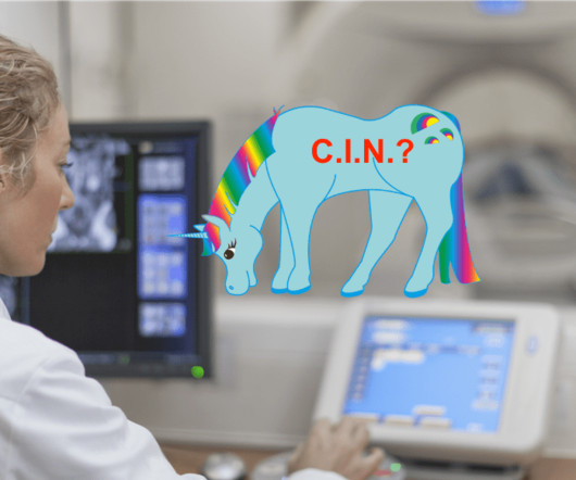
3 A study of CT use trends in the ED has shown increasing use of CTs by almost 60% from 2005 to 2013. CT scans have become an integral part of the ED diagnostic process, serving as a diagnostic tool to aid in determining appropriate disposition, risk assessment, and guiding admission decisions.
Philadelphia: Elsevier, 2013 (Ch) 26: p. et al, Emergency Medicine Clinical Essentials ed 2. 209-225 Walker R, Adhikari S.: Eye Emergencies, in Tintinalli J et al (eds): Tintinalli’s Emergency Medicine: A Comprehensive Study Guide, Seventh Edition New York City: McGraw-Hill 2016 (Ch) 241 Guluma K, Lee JE.
Moreover, the research which appears to confirm this idea was indeed in relation to the circumflex, but they did not study Occlusion ; rather, they studied asymptomatic coronary disease. I showed conclusively that this is a common finding in normal ECGs, though it is more common in LAD Occlusion than in norml variant STE.
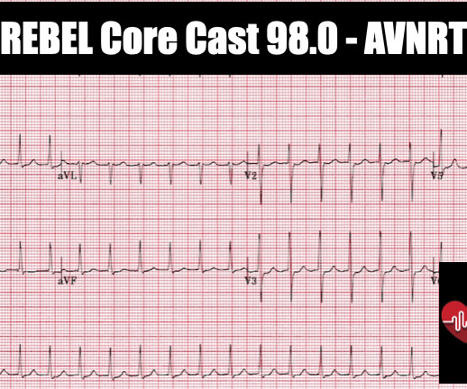
Resources REBEL EM: SVT with Aberrancy Versus VT Amal Mattu’s ECG Case of the Week: August 26th, 2013 ALiEM: Tricks of the Trade: Valsalva Maneuver By Using a 10cc Syringe Larry Mellick: Treating SVT with Adensoine ALiEM: Trick of the Trade: Combining Adenosine with the Flush References Brugada P et al.
Along the way to acquiring more experience in recognizing the ECG findings of acute coronary occlusion — is incorporation of a number of KEY ECG Features into one's clinical acumen. Although this is not a common phenomenon — You will see it on occasion ( See the July 24, 2013 post , among others — in Dr. Smith's ECG Blog ).
Int J Cardiol 2013 2. Identifying patients with low risk for acute coronary syndrome without troponin testing: validation of the HEAR score. High-sensitivity cardiac troponin I at presentation in patients with suspected acute coronary syndrome: a cohort study. Backus BE, Six AJ, Kelder JC, et al. Shin YS, Ahn S, Kim YJ.
This " imbalance of precordial T waves" is not seen very often — and in the “right” clinical setting, has been associated with recent OMI from a LCx culprit artery ( See Manno et al: JACC 1:1213, 1983 — and the July 17, 2013 post by Salim Rezaie in ALiEM ).
Posterior STEMI" may not even technically exist according to the current (2013) ACC/AHA STEMI guidelines, as it is not described as a "STEMI equivalent" and the only relevant statement in the guidelines is: "In addition, ST depression in 2 precordial leads (V1-V4) may indicate transmural posterior injury." Comment from K. What is "STEMI"?
PEARL #2 — As emphasized by Dr. Smith in the July 24, 2013 post in Dr. Smith’s ECG Blog — the ST-T wave appearance in repolarization variants may be dynamic ! The angiogram showed completely normal vessels ( No evidence of coronary disease! ). As a result — I thought cardiac cath was clearly indicated to clarify the clinical picture.
The acute coronary syndrome work-up is negative but she is Well’s high and needs a CTPA to rule-out a pulmonary embolism. Background: There has been a huge increase in the number of CT scans performed with more than 75 million CT scans performed in the US in 2013. Case: A 64-year-old woman with type-2 diabetes.
Post by Smith and Meyers Sam Ghali ( [link] ) just asked me (Smith): "Steve, do left main coronary artery *occlusions* (actual ones with transmural ischemia) have ST Depression or ST Elevation in aVR?" Furthermore, among 35 patients with acute left main coronary artery occlusion, 9 presented with RBBB (mostly with LAFB) on the admission ECG.
STREAM-2: Half-Dose Tenecteplase or Primary Percutaneous Coronary Intervention in Older Patients With ST-Segment-Elevation Myocardial Infarction: A Randomized, Open-Label Trial. Based on this, the authors did a literature review and found that there is an increasing rate of ICH and major non-intracranial bleeding starting at ≈60 years of age.
Am J Cardiol 2013. A Randomized Trial of the Optimum Duration of Acoustic Pulse Thrombolysis Procedure in Acute Intermediate-Reisk Pulmonary Embolism: The OPTALYSE PE Trial. JACC Cardiovasc Interv 2018. PMID: 30025734 Sharifi M et al. Moderate Pulmonary Embolism Treated with thrombolysis (from the “MOPETT” Trial). Clin Exp Emerg Med 2023.
Electrocardiographic Diagnosis of Acute Coronary Occlusion Myocardial Infarction in Ventricular Paced Rhythm Using the Modified Sgarbossa Criteria. Immediate ECG diagnosis of an acute coronary lesion resulting in occlusion myocardial infarction is critical in the modern reperfusion era. Annals of Emergency Medicine 2021.
Background: Coronary artery disease can result in hibernating myocardium (chronic myocardial contractile dysfunction) due to ischemia. The theory is that there is reduced coronary blood flow and increased myocardial demand resulting in impaired contractility. Paper: Perera D et al. OMT: 38.0% HR 0.99; 95% CI 0.78 to 1.27; p = 0.96
Rather it is due to coronary insufficiency due to a tight left main or 3-vessel disease with inadequate coronary flow. Fourteen (16%) of the 86 patients who had CT also had immediate cardiac catheterization for acute ischemic changes; 7 of which had primary percutaneous coronary intervention (PCI).
The patient proceeded to cath where all coronaries were described as normal with no evidence of any CAD, spasm, or any other abnormality. PM Cardio digitized version. QOH Interpretation: The initial troponin I (older generation) at the first ED was barely positive at 0.06
Am J Cardiol 12(9):1379-1383; Nov 2013. Among patients with left bundle branch block, T-wave peak to T-wave end time is prolonged in the presence of acute coronary occlusion. Musat DL et al. Correlation of QT Interval Correction Methods During Atrial Fibrillation and Sinus Rhythm. Unfortunately, the topic is very complex.
A 2013 study found that half of young Australians are dissatisfied with school-based sex education. The goal of chest compressions during neonatal resuscitation is to increase cerebral and coronary blood flow with the intention to achieve a return of spontaneous circulation (ROSC). Why does it matter? Sex ed” sucks.
Some of the critical differentials include pulmonary embolism, acute decompensated heart failure, pneumonia, pneumothorax, and acute coronary syndrome. Anginal chest pain, chest heaviness, or evidence of fluid overload suggest acute coronary syndrome or acute decompensated heart failure. Signs and symptoms of systemic infection (e.g.,
Myocardial Infarction with Non-Obstructive Coronary Arteries. Etiologies (list not comprehensive): Coronary Spasm. Provocative testing is very helpful for this Coronary Thrombus with lysis (one must do optical coherence tomography or at least intravascular ultrasound to find thes non-obstructive plaques that ruptured.
Lidocaine had been used for the prevention of VF since the 1960s after coronary care units became a standard setting for the treatment of AMI. Primary VF in this study refers to fibrillation occurring in the absence of shock or pulmonary edema. Mortality rate, when primary VF occurs, is 2 to 4 times greater than when it does not.
Rates of occurrence of PIRP have decreased drastically in the era of percutaneous coronary intervention. Incidence and Prognosis of Pericarditis After ST-Elevation Myocardial Infarction (from the Acute Coronary Syndrome Israeli Survey 2000 to 2013 Registry Database). of patients developed PIRP [ 1 ]. Oliva et al. [ Hammill, S.
The arterial pressure waveform is transduced using the coronary catheter. Normally, the diameter of the coronary artery ostium is much greater than the diameter of the catheter so that catheter engagement does not significantly impair antegrade coronary perfusion. Here is the ECG and arterial waveform during RCA angiography.
We organize all of the trending information in your field so you don't have to. Join 5,000+ users and stay up to date on the latest articles your peers are reading.
You know about us, now we want to get to know you!
Let's personalize your content
Let's get even more personalized
We recognize your account from another site in our network, please click 'Send Email' below to continue with verifying your account and setting a password.


Let's personalize your content