Remove 2019 Remove ACS Remove CAD
article thumbnail

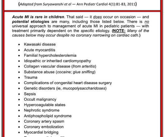
OMI in a pediatric patient? Teenagers do get acute coronary occlusion, so don't automatically dismiss the idea.

Dr. Smith's ECG Blog

He did have a family history notable for early CAD. A final ECG was perfomed on hospital day 2: Persistent ST elevation in the inferior leads with slight reciprocal ST depression in aVL Teaching points - It is essential to consider ACS in all age groups. He denied drug or alcohol use. ng/mL (ULN 16,000 ng/L, mildly elevated CRP of 8.4

Coronary 116
article thumbnail

A 29 year old male with chest pain, ST Elevation, and very elevated troponin T

Dr. Smith's ECG Blog

By Magnus Nossen This ECG is from a young man with no risk factors for CAD, he presented with chest pain. ACS then becomes less likely. Before the lab values returned this patient had a n emergent coronary CT angiogram done that ruled out CAD. How would you assess this ECG? How confident are you in your assessment?

professionals

Sign Up for our Newsletter

This site is protected by reCAPTCHA and the Google Privacy Policy and Terms of Service apply.

article thumbnail

Dark Side of the Moon

EMS 12-Lead

Furthermore, there was no family history of early CAD, MI, or sudden cardiac death. Smith and Meyers found that patients presenting with high-risk ACS and any ST-depression, even less than 1 mm, maximal in leads V1-V4 to be 97% specific for OMI and 96% specific for OMI requiring emergent PCI. [5] 3] Niu, T., 4] Yang, T.,

STEMI 130
article thumbnail

"Pericarditis" strikes again

Dr. Smith's ECG Blog

Written by Pendell Meyers A man in his late 40s with several ACS risk factors presented with a chief complaint of chest pain. The cardiologists felt that the ECG did not represent ACS, and thought it was more likely pericarditis, so they did not take him to the cath lab. His first troponin T then resulted elevated at 0.19

E-9-1-1 52
article thumbnail

A 40 year old man with chest pain since last night

Dr. Smith's ECG Blog

He also had non-acute CAD of the RCA (50%) and LCX (50%). When in doubt, record serial ECGs and watch out for signs of ischemia despite medical management. == Comment by K EN G RAUER, MD ( 7/11/2019 ): == Our thanks to Drs. Cath images: Before intervention. Before intervention with arrows demonstrating the area of occlusion.

STEMI 52
article thumbnail

Upon arrival to the emergency department, a senior emergency physician looked at the ECG and said "Nothing too exciting."

Dr. Smith's ECG Blog

The fire department, who operate at an EMT level in this municipality, arrived before us and administered 324 mg of baby aspirin to the patient due to concern for ACS. Just because you don't see hemodynamically significant CAD on angiogram does not mean it is not OMI. I could have told you this (and did tell you this) without an MRI.

article thumbnail

What will you do for this patient transferred to you who is now asymptomatic?

Dr. Smith's ECG Blog

She did not receive any opioids (which would mask her pain without affecting any underlying ACS). I believe this is by far the most common outcome for this patient around the world in 2019. She also had non-acute CAD of the left main (50%) and LCX (75%). What will you do for this asymptomatic patient??? They opened it.

STEMI 52