Remove 2019 Remove ACS Remove Coronary
article thumbnail

What does the angiogram show? The Echo? The CT coronary angiogram? How do you explain this?

Dr. Smith's ECG Blog

Angiogram No obstructive epicardial coronary artery disease Cannot exclude non-ACS causes of troponin elevation including coronary vasospasm, stress cardiomyopathy, microvascular disease, etc. CORONARY ARTERIES: Exam was not directly tailored for coronary artery evaluation, noting recent diagnostic coronary angiogram.

Coronary 104
article thumbnail

OMI in a pediatric patient? Teenagers do get acute coronary occlusion, so don't automatically dismiss the idea.

Dr. Smith's ECG Blog

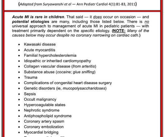
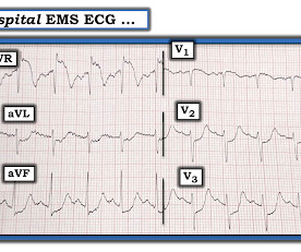
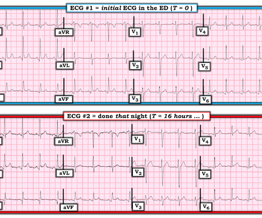
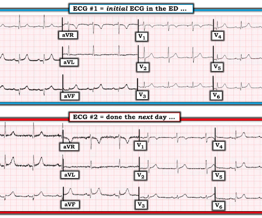
Acute coronary syndrome in a pediatric patient? A final ECG was perfomed on hospital day 2: Persistent ST elevation in the inferior leads with slight reciprocal ST depression in aVL Teaching points - It is essential to consider ACS in all age groups. Ultimately, cardiac cath was done — revealing patent coronary arteries.

Coronary 116
professionals

Sign Up for our Newsletter

This site is protected by reCAPTCHA and the Google Privacy Policy and Terms of Service apply.

article thumbnail

SGEM#344: We Will…We Will Cath You – But should We After An OHCA Without ST Elevations?

The Skeptics' Guide to EM

Acute coronary syndrome (ACS) is responsible for the majority (60%) of all OHCAs in patients. Emergency coronary angiography is reasonable for select (eg, electrically or hemodynamically unstable) adult patients who are comatose after OHCA of suspected cardiac origin but without ST elevation on ECG (Class IIa, LOE B-NR). .

EMR 130
article thumbnail

A 29 year old male with chest pain, ST Elevation, and very elevated troponin T

Dr. Smith's ECG Blog

ACS then becomes less likely. Before the lab values returned this patient had a n emergent coronary CT angiogram done that ruled out CAD. A false positive cath lab activation is also off course acceptable for this diagnosis if you cannot get an emergent coronary CT angiogram. There are no coronary stenoses.

article thumbnail

Concerning EKG with a Non-obstructive angiogram. What happened?

Dr. Smith's ECG Blog

The commonest causes of MINOCA include: atherosclerotic causes such as plaque rupture or erosion with spontaneous thrombolysis, and non-atherosclerotic causes such as coronary vasospasm (sometimes called variant angina or Prinzmetal's angina), coronary embolism or thrombosis, possibly microvascular dysfunction. This is not the case.

E-9-1-1 123
article thumbnail

90 year old with acute chest and epigastric pain, and diffuse ST depression with reciprocal STE in aVR: activate the cath lab?

Dr. Smith's ECG Blog

Smith: If this is ACS (a big if), t his is just the time when one should NOT use "upstream" dual anti-platelet therapy ("upstream" means in the ED before angiography). History sounds concerning for ACS (could be critical stenosis, triple vessel), but differential also includes dissection, GI bleed, etc. Am J Med 2019, 132(5):622-630.

article thumbnail

50 Shades of T

EMS 12-Lead

It should be emphasized here that this is a presentation of high-pretest probability for Acute Coronary Syndrome (ACS). ACS and hyperkalemia both have lethal downstream consequences, so it is imperative for the clinician to acclimate to the presentation, or developing, features of each. ECG's are difficult. 2] Costanzo, L.

ACS 130