Remove 2019 Remove CAD Remove Coronary
article thumbnail

OMI in a pediatric patient? Teenagers do get acute coronary occlusion, so don't automatically dismiss the idea.

Dr. Smith's ECG Blog

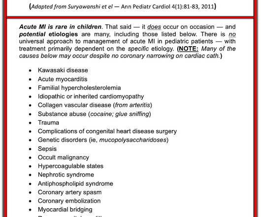
Acute coronary syndrome in a pediatric patient? He did have a family history notable for early CAD. hematological disorder like sickle cell or antiphospholipid syndome, family history of CAD or hypercholesterolemia, prior history of vasculopathies such as Kawasaki Disease, MIS-C, prior cardiac surgery, etc.)

Coronary 116
article thumbnail

A 29 year old male with chest pain, ST Elevation, and very elevated troponin T

Dr. Smith's ECG Blog

By Magnus Nossen This ECG is from a young man with no risk factors for CAD, he presented with chest pain. Before the lab values returned this patient had a n emergent coronary CT angiogram done that ruled out CAD. Each main coronary artery (LAD, RCA and LCx) are shown in separate images. There are no coronary stenoses.

professionals

Sign Up for our Newsletter

This site is protected by reCAPTCHA and the Google Privacy Policy and Terms of Service apply.

article thumbnail

Something Winter This Way Comes

EMS 12-Lead

Otherwise, no admission of CAD, HLD, or family history of sudden cardiac death. A second 12 Lead ECG was recorded: This is a testament to the dynamic nature of coronary thrombosis and thrombolysis. He described the pain as “nagging,” and equally not exacerbated by any kind of movement. But the lesion is still active!

STEMI 130
article thumbnail

Dark Side of the Moon

EMS 12-Lead

Furthermore, there was no family history of early CAD, MI, or sudden cardiac death. Cardiology admitted him for observation with plans for next-day coronary angiogram. He reported to EMS a medical history of GERD only. The physical exam was unremarkable for diaphoresis or pallor, and he denied any episodes of vomiting. 1] Driver, B.

STEMI 130
article thumbnail

What would you do with acute chest pain and this ECG? You might see what the Queen thinks.

Dr. Smith's ECG Blog

The ED provider ordered a coronary CT scan to assess the patient for CAD. The patient was taken emergently to the cath lab for a pericardiocentesis instead of a coronary angiogram. Three months prior to this presentation, he received a pacemaker for severe bradycardia and syncope due to sinus node dysfunction.

STEMI 63
article thumbnail

"Pericarditis" strikes again

Dr. Smith's ECG Blog

They found non-obstructive CAD, with only a 20% stenosis of OM2 and 10% RCA. That said — I illustrate HOW this RATIO is arrived at in Figure-2 , which I have adapted from the 3/16/2019 post in Life-In-The-Fast-Lane. A repeat ECG was performed and cardiology was re-consulted: Roughly unchanged. No acute culprit.

E-9-1-1 52
article thumbnail

A 40 year old man with chest pain since last night

Dr. Smith's ECG Blog

He also had non-acute CAD of the RCA (50%) and LCX (50%). When in doubt, record serial ECGs and watch out for signs of ischemia despite medical management. == Comment by K EN G RAUER, MD ( 7/11/2019 ): == Our thanks to Drs. This is a h igher - p revalence H istory for acute coronary disease. Cath images: Before intervention.

STEMI 52