Remove 2019 Remove Coronary Remove E-9-1-1
article thumbnail

What does the angiogram show? The Echo? The CT coronary angiogram? How do you explain this?

Dr. Smith's ECG Blog

Angiogram No obstructive epicardial coronary artery disease Cannot exclude non-ACS causes of troponin elevation including coronary vasospasm, stress cardiomyopathy, microvascular disease, etc. CORONARY ARTERIES: Exam was not directly tailored for coronary artery evaluation, noting recent diagnostic coronary angiogram.

Coronary 104
article thumbnail

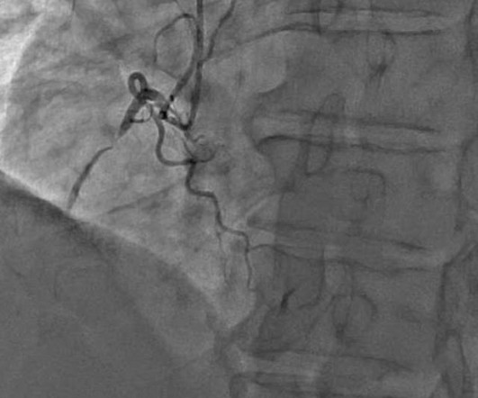
Concerning EKG with a Non-obstructive angiogram. What happened?

Dr. Smith's ECG Blog

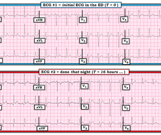
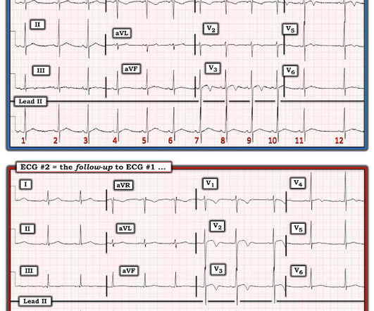
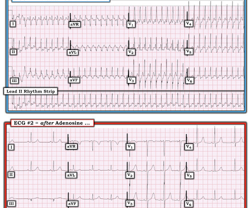
link] A 62 year old man with a history of hypertension, type 2 diabetes mellitus, and carotid artery stenosis called 911 at 9:30 in the morning with complaint of chest pain. Challenge QUESTION: The relative change in T-QRS-D is not the only thing that changes during period of time that passed between recording of the 2 ECGs shown in Figure-1.

E-9-1-1 123
professionals

Sign Up for our Newsletter

This site is protected by reCAPTCHA and the Google Privacy Policy and Terms of Service apply.

article thumbnail

What Lies Beneath

EMS 12-Lead

Question 1: What is the rhythm? Beat 1 : Sinus, narrow QRS complex. The assumption is that a premature complex discharged prior to Beat 1, which prolonged its respective refractory period in the same manner as Beat 5. The coronary angiogram revealed no critical stenosis, or acute plaque ulceration.

E-9-1-1 130
article thumbnail

How important are old ECGs in Non-obvious cases of potential OMI?

Dr. Smith's ECG Blog

We who know ischemic ECGs know that really when T-wave inversion is specific for coronary thrombosis that it indicates reperfusion of the artery, not active occlusion. Learning Point: 1. For examples of such exceptions — See My Comment in the January 9, 2019 — August 22, 2020 — and June 30, 2023 posts in Dr. Smith's ECG Blog ).

E-9-1-1 111
article thumbnail

Teenager with chest pain and slightly elevated troponin. What happens then?

Dr. Smith's ECG Blog

The pain is described as located in the midsternal area, radiating to the right arm, described as 8-9/10 and worse with deep inspirations. In the evening, he became diaphoretic and complained of 9/10 continuous chest pain. If there is NO WMA, then it is not coronary occlusion, but if there IS one, then you still don't have a diagnosis.

E-9-1-1 52
article thumbnail

A man in his 50s with chest pain

Dr. Smith's ECG Blog

He had episodes of chest pain off and on all night, until about 1 hour prior to arrival when the pain became constant, crushing, 10/10 chest pain that radiated to both arms. Proven STEMI has an open artery in 19% to 36% of cases, depending on whether it is TIMI −1, −2, or −3 flow. 25] Stone et al found that 72% have TIMI 0 or 1 flow.

E-9-1-1 52
article thumbnail

Extreme shock and cardiac arrest in COVID patient

Dr. Smith's ECG Blog

And so it is wise to look at the coronary arteries. Here they are: Learning Points: 1. This ECG certainly looks like myocarditis, and was due to myocarditis, but missing acute coronary occlusion is not acceptable. In acute MI, the T-wave is large, and the T/ST ratio is high. This is much more typical of myocarditis.

E-9-1-1 52