Remove 2019 Remove Coronary Remove ED
article thumbnail

What does the angiogram show? The Echo? The CT coronary angiogram? How do you explain this?

Dr. Smith's ECG Blog

[link] Case continued She arrived in the ED and here is the first ED ECG. Angiogram No obstructive epicardial coronary artery disease Cannot exclude non-ACS causes of troponin elevation including coronary vasospasm, stress cardiomyopathy, microvascular disease, etc. Detailed coronary artery evaluation not performed.

Coronary 104
article thumbnail

OMI in a pediatric patient? Teenagers do get acute coronary occlusion, so don't automatically dismiss the idea.

Dr. Smith's ECG Blog

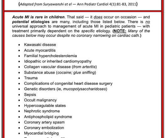
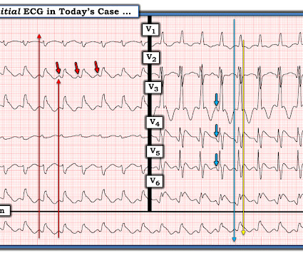
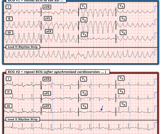
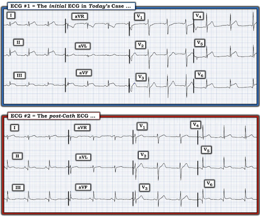
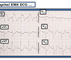
Acute coronary syndrome in a pediatric patient? An ECG was perfomed on arrival to our ED: NSR with ST elevation II,III, aVF with reciprocal depression in aVL Would you refer this pediatric patient for emergent PCI? Ultimately, cardiac cath was done — revealing patent coronary arteries. mg/L and a normal WBC of 8.8.

Coronary 116
professionals

Sign Up for our Newsletter

This site is protected by reCAPTCHA and the Google Privacy Policy and Terms of Service apply.

article thumbnail

SGEM#344: We Will…We Will Cath You – But should We After An OHCA Without ST Elevations?

The Skeptics' Guide to EM

He is interested and experienced in healthcare informatics, previously worked with ED-directed EMR design, and is involved in the New York City Health and Hospitals Healthcare Administration Scholars Program (HASP). She arrives in the emergency department (ED) with decreased level of consciousness and shock.

EMR 130
article thumbnail

Something Winter This Way Comes

EMS 12-Lead

A second 12 Lead ECG was recorded: This is a testament to the dynamic nature of coronary thrombosis and thrombolysis. Here the ST segments are not so deep, nor are the T waves so wide and bulky, because of improved coronary flow at the level of the occlusion. But the lesion is still active! However, when the Troponin I returned 8.4

STEMI 130
article thumbnail

Hypertrophic Cardiomyopathy

EMS 12-Lead

Additional architectural changes include systolic anterior motion of the mitral valve, endothelial dysfunction at the level of the coronary arterial bed, and ventricular diastolic dysfunction. This worried the crew of potential acute coronary syndrome and STEMI was activated pre-hospital. Below is the initial ED ECG.

Coronary 130
article thumbnail

Concerning EKG with a Non-obstructive angiogram. What happened?

Dr. Smith's ECG Blog

The commonest causes of MINOCA include: atherosclerotic causes such as plaque rupture or erosion with spontaneous thrombolysis, and non-atherosclerotic causes such as coronary vasospasm (sometimes called variant angina or Prinzmetal's angina), coronary embolism or thrombosis, possibly microvascular dysfunction. This is not the case.

E-9-1-1 123
article thumbnail

A man in his 50s with acute chest pain who is lucky to still be alive.

Dr. Smith's ECG Blog

Below is the version standardized by PM Cardio app Meyers interpretation: Findings are specific for posterior (and also likely inferior) wall transmural acute infarction, most likely due to acute coronary occlusion (OMI). Clinical Cardiology 2019. In this case, the EMS provider was routed to the urgent care facility.

STEMI 97