Remove 2019 Remove Defibrillator Remove ED
article thumbnail

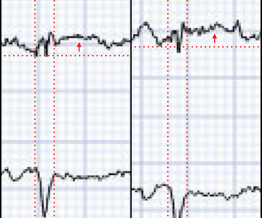
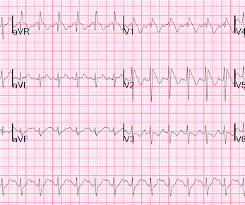
Guidelines would (erroneously) say that this patient who was defibrillated and resuscitated does not need emergent angiography

Dr. Smith's ECG Blog

A patient had a cardiac arrest with ventricular fibrillation and was successfully defibrillated. Here was his initial ED ECG: Formal interpretation by interventional cardiologist: There is "Non-diagnostic" ST Elevation in V2-V4 and aVL. Lemkes JS, Janssens GN, van der Hoeven NW, et al.

article thumbnail

SGEM#344: We Will…We Will Cath You – But should We After An OHCA Without ST Elevations?

The Skeptics' Guide to EM

He is interested and experienced in healthcare informatics, previously worked with ED-directed EMR design, and is involved in the New York City Health and Hospitals Healthcare Administration Scholars Program (HASP). She arrives in the emergency department (ED) with decreased level of consciousness and shock.

EMR 130
professionals

Sign Up for our Newsletter

This site is protected by reCAPTCHA and the Google Privacy Policy and Terms of Service apply.

article thumbnail

What the Emergency Clinician Must Know about Remote Monitors in Heart Failure

EMDocs

He has been taking increased doses of torsemide without improvement in his symptoms and was directed to the ED for inpatient therapy. Now, a remote outpatient pulmonary arterial pressure monitor and other heart failure detection devices (Table 1) are available that you may begin to see in your ED.

article thumbnail

Hypothermic Cardiac Arrest: Pearls and Pitfalls

EMDocs

Louis) // Reviewed by: Alex Koyfman, MD (@EMHighAK); Brit Long, MD (@long_brit) Case You are working in the trauma/critical care pod of your emergency department (ED). His blood sugar was normal en route to the ED, and his initial rhythm on the cardiac monitor was asystole. It is unclear how long he was down.

article thumbnail

A man in his 50s with acute chest pain who is lucky to still be alive.

Dr. Smith's ECG Blog

In this medical system, the EMS provider can then be routed to the ED or to a type of urgent care facility that is open 24 hrs/day and staffed by a primary care provider. Clinical Cardiology 2019. In this case, the EMS provider was routed to the urgent care facility. The LCX was noted to be "subtotally stenosed" at the time of cath.

STEMI 97
article thumbnail

What Lies Beneath

EMS 12-Lead

We can, therefore, put down the defibrillation pads, set aside the amiodarone, and look further at the ECG. Chou’s Electrocardiography in Clinical Practice (6th ed). Paradoxically, though, the third green arrow identifies a QRS that is more narrow than the RBBB complexes surrounding it. Pacing Clin Electrophysiol., 40, 1234-41.

E-9-1-1 130
article thumbnail

Noisy, low amplitude ECG in a patient with chest pain

Dr. Smith's ECG Blog

Rhythm B: This patient was seen in the ED — and thought to be in AFlutter with 4:1 AV conduction. Rhythm C: This telemetry strip from an older adult was initially thought to need defibrillation. The September 27, 2019 post — for the Rowlands & Moore article with the above-noted formulas for recognizing the “culprit” extremity.