Remove 2019 Remove Defibrillator Remove STEMI
article thumbnail

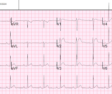
Guidelines would (erroneously) say that this patient who was defibrillated and resuscitated does not need emergent angiography

Dr. Smith's ECG Blog

A patient had a cardiac arrest with ventricular fibrillation and was successfully defibrillated. COACT: The COACT trial was fatally flawed, and because of it, many cardiologists are convinced that if there are no STEMI criteria, the patient does not need to go to the cath lab. Lemkes JS, Janssens GN, van der Hoeven NW, et al.

article thumbnail

A man in his 50s with acute chest pain who is lucky to still be alive.

Dr. Smith's ECG Blog

You can subscribe for news and early access (via participating in our studies) to the Queen of Hearts here: [link] queen-form This EMS ECG was transmitted to the nearby Emergency Department where it was remotely reviewed by a physician, who interpreted it as normal, or at least without any features of ischemia or STEMI.

STEMI 97
professionals

Sign Up for our Newsletter

This site is protected by reCAPTCHA and the Google Privacy Policy and Terms of Service apply.

article thumbnail

SGEM#344: We Will…We Will Cath You – But should We After An OHCA Without ST Elevations?

The Skeptics' Guide to EM

Defibrillation is the treatment of choice in these cases but does not often result in sustained ROSC ( Kudenchuk et al 2006). There is evidence that taking those patients with ROSC and EKG showing STEMI directly for angiography +/- angioplasty is associated with positive patient-oriented outcomes.

EMR 130
article thumbnail

What are treatment options for this rhythm, when all else fails?

Dr. Smith's ECG Blog

The ECG shows obvious STEMI(+) OMI due to probable proximal LAD occlusion. He required multiple defibrillations within a period of a few hours. This time, the arrhythmia did not spontaneously terminate — but rather degenerated to VFib, requiring defibrillation. The below ECG was recorded. What do you think?

article thumbnail

A Middle-Aged male with Chest Pain and an Unusual ECG

Dr. Smith's ECG Blog

If it is STEMI, it would have to be RBBB with STEMI. Cardioversion/defibrillation. But — one of the causes of Brugada Phenocopy is acute infarction — so I didn’t know how to distinguish between a preexisting Brugada-1 ECG pattern vs a Brugada ECG pattern developing as a result of acute ongoing anterior STEMI. Hypothermia.

STEMI 52
article thumbnail

Two 70 year olds with chest pain, and 3 pitfalls of the STEMI paradigm

Dr. Smith's ECG Blog

There’s inferior ST depression which is reciprocal to subtle lateral convex ST elevation, and the precordial T waves are subtly hyperacute – all concerning for STEMI(-)OMI of proximal LAD. There’s ST elevation I/aVL/V2 that meet STEMI criteria. This is obvious STEMI(+)OMI of proximal LAD. Non-STEMI or STEMI(-)OMI?

STEMI 52
article thumbnail

Interventionalist at the Receiving Hospital: "No STEMI, no cath. I do not accept the transfer."

Dr. Smith's ECG Blog

Are Some Cardiologists Really Limited by Strict Adherence to STEMI millimeter criteria? He was found in ventricular fibrillation and defibrillated, then brought to a local ED which does not have a cath lab. This is the response he got: Interventionist: "No STEMI, no cath. It is a STEMI equivalent.

STEMI 52