This site uses cookies to improve your experience. To help us insure we adhere to various privacy regulations, please select your country/region of residence. If you do not select a country, we will assume you are from the United States. Select your Cookie Settings or view our Privacy Policy and Terms of Use.
Cookie Settings
Cookies and similar technologies are used on this website for proper function of the website, for tracking performance analytics and for marketing purposes. We and some of our third-party providers may use cookie data for various purposes. Please review the cookie settings below and choose your preference.
Used for the proper function of the website
Used for monitoring website traffic and interactions
Cookie Settings
Cookies and similar technologies are used on this website for proper function of the website, for tracking performance analytics and for marketing purposes. We and some of our third-party providers may use cookie data for various purposes. Please review the cookie settings below and choose your preference.
Strictly Necessary: Used for the proper function of the website
Performance/Analytics: Used for monitoring website traffic and interactions
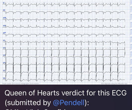
The 2022 American College of Cardiology (ACC) pathway provides timely guidance [1]. Applying the 2022 ACC guideline Before delving into the specifics of the hs-cTn pathways, start with the ECG. The ACC 2022 pathway has a section dedicated to ECGs in ischemia [1], and FOAMcast has a great visual summary.
What Your Gut Says: The patient has a tachydysrhythmia which may be the presentation of acute coronary syndrome (ACS) even though the patient has no ischemic symptoms. Up to 80% of patients will have at least one troponin sent ( Gabrielli 2022 ). SVT is not a presenting dysrhythmia consistent w/ ACS. Cardiol Rev.
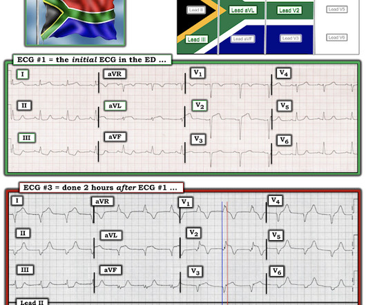
Review of the 2 ECGs in today's case is insightful ( Figure-1 ): The initial ECG shows sinus rhythm, LAHB and meets Peguero Criteria for LVH ( See My Comment in the August 15, 2022 post of Dr. Smith's ECG Blog for more on LVH criteria ). Nor was there a challenge to look for coronary spasm.
Other causes of sickling: acidosis, dehydration, inflammation, infection, fever, and blood stasis Sickling leads to vascular occlusion, end-organ ischemia, and decreased RBC lifespan, which, in turn, leads to pain crisis, acute anemia, sequestration, infection, and acute chest syndrome (ACS.) Each episode of ACS has a 9% mortality rate.
Date: June 30th, 2022 Reference: McGinnis et al. AEM June 2022. Date: June 30th, 2022 Reference: McGinnis et al. AEM June 2022. If we thought about ACS, we brought them in. AEM June 2022. Major adverse cardiac event rates in moderate-risk patients: Does prior coronary disease matter?
Sickling leads to vascular occlusion, end-organ ischemia, and decreased RBC lifespan, which, in turn, leads to pain crisis, acute anemia, sequestration, infection, and acute chest syndrome (ACS). ACS is lung injury due to vaso-occlusion in the pulmonary vasculature; many with ACS will have a concomitant vaso-occlusive pain crisis.
The person I was texting knows implicitly based on our experience together that I mean "Definite posterior OMI, assuming the patient's clinical presentation is consistent with ACS." The patient was a middle-aged female who had acute chest pain of approximately 6 hours duration. The pain was still active at the time of evaluation.
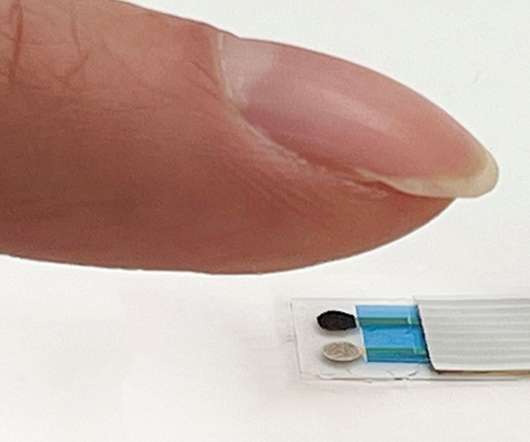
” The researchers presented their technology at the 2022 fall meeting of the American Chemical Society (ACS). Via: ACS. . “We already interact with a lot of touch-based electronics, such as smart phones and keyboards, so this sensor could integrate seamlessly into daily life.”
Date: May 24th, 2022 Reference: Broder et al. Date: May 24th, 2022 Reference: Broder et al. AEM May 2022 Guest Skeptic: Dr. Justin Morgenstern is an emergency physician and the creator of the #FOAMed project called First10EM.com Case: A 33-year-old male presents to the emergency department (ED) complaining of abdominal pain.
As a result, even before looking at this patient's initial ECG — he falls into a high -prevalence likelihood group for ACS ( for an A cute C oronary S yndrome ). We therefore need to assume and rule "out" ACS — more than having to rule it "in". The "onus of proof" remains on us as medical providers to objectively rule out ACS.
An expert committee appraised the evidence behind recommendations to avoid imaging to inform the 2022 NICE guidance. Published 2022 Jan 13. doi:10.1177/0883073818786086 Young AC, Costanzi JB, Mohr PD, Forbes WS. Paediatr Child Health. 2021;26(1):50-57 NICE. NICE guideline NG217. Accessed online at [link] NICE. Front Pediatr.
Smith : As Willy states, ACS with persistent symptoms is a guideline recommended indication for <2 hour angio (both ACC/AHA and ESC). The ESC states that patients with suspected ACS should go to the cath lab in <2 hours "regardless of ECG or biomarker evidence of MI!!" See this case: A man his 50s with chest pain.
But because the patient had no chest pain or shortness of breath, it was not deemed to be from ACS. They were less likely to have STEMI on ECG, and more likely to be initially diagnosed as non-ACS. Dialysis patients have a high rate of ACS without chest pain and high rate of delayed diagnosis and delayed reperfusion 2.
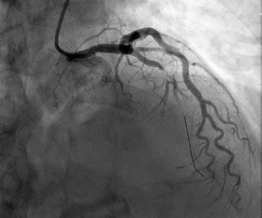
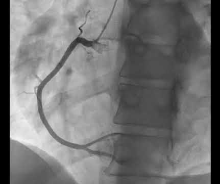
Angiogram No obstructive epicardial coronary artery disease Cannot exclude non-ACS causes of troponin elevation including coronary vasospasm, stress cardiomyopathy, microvascular disease, etc. Most studies examine undifferentiated ACS cohorts, with only a handful providing separate data. References: 1.
Morley 2022) This myth is: BUSTED. References: Ross RK, Kinlaw AC, Herzog MM, Funk MJ, Gerber JS. Symptoms resolved within 24 hours of withdrawal of the medication. This is the 8 th case report which has identified fluoroquinolone-associated peripheral neuropathy in a child. Nobody wants a pseudomonas superbug becoming prevalent!
In our opinion it should not be given in ACS unless you are committed to the cath lab. Learning Point: Any NSTEMI patient with active ongoing ACS symptoms refractory to medical management is supposed to go to the cath lab within 2 hours if available, per all guidelines in world, regardless of ECG findings. Published 2022 Feb 20.
Ischemia from ACS causing the chest discomfort, with VT another consequence (or coincidence)? Cardioversion will address the rhythm problem immediately, also if the chest discomfort subsides when SR is restored, ischemia from ACS becomes much less likely. In either case, prompt cardioversion is indicated.
Non-STEMI guidelines call for “urgent/immediate invasive strategy is indicated in patients with NSTE-ACS who have refractory angina or hemodynamic or electrical instability,” regardless of ECG findings.[1] Clin Cardiol 2022 4. 1] European guidelines add "regardless of biomarkers". But only 6.4% Int J Cardiol 2024 3. Lupu et al.
And now this finding is even formally endorsed as a "STEMI equivalent" in the 2022 ACC guidelines!!! See this study showing an association between morphine and mortality in ACS: Use of Morphine in ACS is independently associated with mortality, at odds ratio of 1.4. They are simply Hyperacute T-waves with depressed ST takeoff.
Thus, this does NOT meet STEMI criteria (though, as of 2022, it is a formal "STEMI equivalent", assuming everyone agrees that this is de Winter morphology, for which there is currently no objective definition). Also, if you use the LAD OMI formula : QT = 420, RAV4 = 5 mm, QRSV2 = 6 mm, STE60V3 = 2.5 mm, the value is 22.2 (LAD
Smith: If this is ACS (a big if), t his is just the time when one should NOT use "upstream" dual anti-platelet therapy ("upstream" means in the ED before angiography). History sounds concerning for ACS (could be critical stenosis, triple vessel), but differential also includes dissection, GI bleed, etc. Anything more on history?
NOTE: For more on “My Take” regarding a historical perspective, including current clinical relevance of recognizing Wellens' Syndrome — See My Comment at the bottom of the page of the August 12, 2022 post. How to Check Your Findings. To Emphasize — I would not be at all sure from the subtle findings in ECG #1 that there was ongoing LAD OMI.
2022 Aug;58:223-228. Wilkerson RG, Ogunbodede AC. High risk and low prevalence diseases: Eclampsia. Am J Emerg Med. Fishel Bartal M, Sibai BM. Eclampsia in the 21st century. American journal of obstetrics and gynecology. 2022;226(2S):S1237-S1253. Gestational Hypertension and Preeclampsia: ACOG Practice Bulletin, Number 222.
2023 Apr 20:fetalneonatal-2022-324835. 2023 Apr 4:archdischild-2022-325281. De Alwis AC, et al. Effect of initial and subsequent mask applications on breathing and heart rate in preterm infants at birth. Kuypers KLAM, et al. Arch Dis Child Fetal Neonatal Ed. Hegeman EM, et al. Pediatr Infect Dis J. 2023 Apr 18. Arch Dis Child.
MOREVER, the morphology of the TWI is just not right for ACS. showed that, when T-waves are inverted in precordial leads, if they are also inverted in lead III and V1, then pulmonary embolism is far more likely than ACS. Figure-2: ECG findings associated of acute PE ( reproduced from My Comment in the March 28, 2022 post ).
ACS omega , 7 (24), pp.20441-20456. Algeo, Thomas J., and Jun Shen. “Theory and classification of mass extinction causation.” ” National Science Review 11.1 2024): nwad237. Olynyk, J.K. and Ramm, G.A., Hemochromatosis. New England Journal of Medicine , 387 (23), pp.2159-2170. Barton, James C. 2013): 403-412. . Girelli, D.,
The NIHSS cutoff that predicts outcomes is 4 points higher in AC compared with PC infarctions. Updated 2022 Dec 22]. Updated 2022 Oct 15]. doi:10.1136/neurintsurg-2022-018715 Jorgensen H, Nakayama H, Raaschou H, Larsen K, Hubbe P, Olsen T. NIHSS does have limitations when applied to posterior circulation (PC) strokes.
Lactate Troponin Could this be ACS or myocarditis? Treasure Island (FL): StatPearls Publishing; April 30, 2022. Tox Ethanol ASA/APAP Serum Osm On repeat exam is there any more concern for toxic ingestion we may consider adding on APAP/ASA or looking for toxic alcohols. Is there additional imaging indicated? Sinus Tachycardia.
If this is ACS with Aslanger's pattern , the ST depression vector of subendocardial ischemia (due to simultaneous 3 vessel or left main ACS) is directed toward lead II (inferior and lateral). Also, ACS does not cause hypoxemia out of proportion to hemodynamic compromise, as PE can.
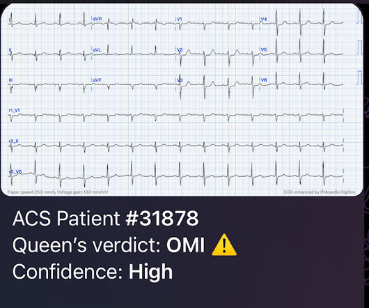
Remember: these findings above are included as STEMI equivalent findings in the 2022 ACC Expert Consensus Decision Pathway on ACS Patients in the ED. Learning Points: To take optimal care of ACS patients, the initial providers who see the patient (hopefully assisted by AI soon) must be able to recognize subtle OMI findings.
The patient was thought to have low likelihood of ACS, and cardiology recommended repeat troponin, urine drug testing, and echocardiogram. At that point, cardiology elected to treat for ACS. NOTE: For review of 20 cases of "Swirl" vs Swirl "Look-Alikes" — Check out the October 15, 2022 post in Dr. Smith's ECG Blog.
Considering hyperacute T-waves have been accepted as STEMI equivalents, it is possible that pseudonormalization could gain more recognition as an indicator of ACS. 2022 Nov, 80 (20) 1925–1960. 5 Studies looking at this phenomenon in the emergency department setting for patients presenting with chest pain are lacking. J Am Coll Cardiol.
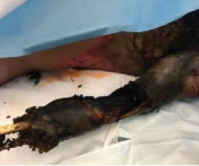
Current can be alternating current (AC) or direct current (DC) with AC typically more dangerous as it is more likely to cause tetanic contractions and increase contact time with the electrical source. 2,3,5 Except for laundry or electrical car outlets (240 V AC), all U.S. household outlets are rated at 120 V AC.
For examples of this phenomenon — See My Comment in the February 14, 2018 — July 21, 2020 — and December 22, 2022 posts in Dr. Smith's ECG Blog ). The emergency physician was skeptical and believed the ECG to be a mimic, a false positive. So they looked into the patient's chart.
This is now further confirmation of ACS. The ACC/AHA guidelines mandate less than 2 hours cath for patients with ACS with refractory pain, pulmonary edema, or electrical or hemodynamic instability. Another ECG was recorded at 160 minutes: There is evolution, with worsening of ischemia.
Clin Cardiol 2022; [link] Labs included: hsTnI 156 ng/L, Hb 12 g/dL, WBC 12x10^9/L, Cr. Smith comment: We have shown that use of opiates is associated with worse outcomes in ACS: Bracey, A. Opioids in ACS may reduce the pain score, but do not provide reperfusion for ongoing ACS. Lupu L, et al. mg/dL, K 3.5
The fire department, who operate at an EMT level in this municipality, arrived before us and administered 324 mg of baby aspirin to the patient due to concern for ACS. Most studies examine undifferentiated ACS cohorts, with only a handful providing separate data. Reference on Troponins: Xenogiannis I, Vemmou E, Nikolakopoulos I, et al.
2022 Dec;57(12):986-993. Joint statement from the American College of Surgeons Committee on Trauma (ACS COT) and the American College of Emergency Physicians (ACEP) regarding the clinical use of Resuscitative Endovascular Balloon Occlusion of the Aorta (REBOA). Trials 23, 384 (2022). J Pediatr Surg. Trauma Surg Acute Care Open.
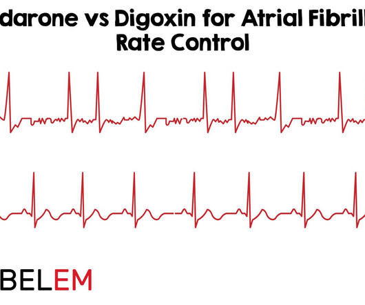
Sepsis, hyperthyroidism, dehydration, heart failure, ACS, etc). 2022 Sep 7. However, digoxin is known for its rate-control properties and its direct vagal effect on the atrioventricular node. Paper: Mason JM, et al. Amiodarone versus digoxin for acute rate control of atrial fibrillation in the emergency department. Am J Emerg Med.
The ECG is diagnostic for acute transmural infarction of the anterior and lateral walls, with LAD OMI being the most likely cause (which has various potential etiologies for the actual cause of the acute coronary artery occlusion, the most common of which is of course type 1 ACS, plaque rupture with thrombotic occlusion).
He had no symptoms of ACS. His HEAR score (before troponin resulted) was documented at 3, with documentation stating "low suspicion for ACS." A troponin this high in a patient with no known chronic troponin elevation, and active acute ACS symptoms, has a very high likelihood of type 1 ACS regardless of the ECG.
You must understand this and the dynamic nature of ACS to provide excellent care for such patients. Comment by K EN G RAUER, MD ( 12/12 /2022 ): = I will summarize in 4 words the important message conveyed by Dr. Meyers in today's post = "Be Aware of Pseudo-Normalization!"
In the December 5, 2022 post of Dr. Smith's ECG Blog — We show 4 additional cases of this pulse-tap artifact. Finally, as I discuss in My Comment in the August 26, 2022 post ( which applies the electrophysiologic principles of Rowlands & Moore: J. This is no longer the case! Why are the smallest artifactual deflections GREEN ?
We organize all of the trending information in your field so you don't have to. Join 5,000+ users and stay up to date on the latest articles your peers are reading.
You know about us, now we want to get to know you!
Let's personalize your content
Let's get even more personalized
We recognize your account from another site in our network, please click 'Send Email' below to continue with verifying your account and setting a password.


Let's personalize your content