This site uses cookies to improve your experience. To help us insure we adhere to various privacy regulations, please select your country/region of residence. If you do not select a country, we will assume you are from the United States. Select your Cookie Settings or view our Privacy Policy and Terms of Use.
Cookie Settings
Cookies and similar technologies are used on this website for proper function of the website, for tracking performance analytics and for marketing purposes. We and some of our third-party providers may use cookie data for various purposes. Please review the cookie settings below and choose your preference.
Used for the proper function of the website
Used for monitoring website traffic and interactions
Cookie Settings
Cookies and similar technologies are used on this website for proper function of the website, for tracking performance analytics and for marketing purposes. We and some of our third-party providers may use cookie data for various purposes. Please review the cookie settings below and choose your preference.
Strictly Necessary: Used for the proper function of the website
Performance/Analytics: Used for monitoring website traffic and interactions
The 2022 American College of Cardiology (ACC) pathway provides timely guidance [1]. Intermediate-risk patients may be further stratified based on recent stress testing or coronary angiogram findings plus a modified HEART or Emergency Department Assessment of Chest Pain (EDACS) score. Time to know your hs-cTn better.
Studies such as those by Moise et al 14 and Ellis et al 39 have shown that the relative risk of developing an acute myocardial infarction in the territory supplied by an artery with a 70%. years, with the interval as long as 12 or 18 years in some studies. Unfortunately, vascular remodeling is variable and inconsistent.
Date: June 30th, 2022 Reference: McGinnis et al. Major adverse cardiac event rates in moderate-risk patients: Does prior coronary disease matter? AEM June 2022. Date: June 30th, 2022 Reference: McGinnis et al. Major adverse cardiac event rates in moderate-risk patients: Does prior coronary disease matter?
” – Musings of an American ED resident in July 2022 when US healthcare was affected simultaneously by supply chain issues from GE Healthcare (contrast media) and Abbott Laboratories (Similac baby formula). A baby formula milk shortage for adults.” 11 Table 1.
What Your Gut Says: The patient has a tachydysrhythmia which may be the presentation of acute coronary syndrome (ACS) even though the patient has no ischemic symptoms. Up to 80% of patients will have at least one troponin sent ( Gabrielli 2022 ). Type 2: MI secondary to ischaemia, but not related to coronary atherosclerosis.
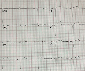
1] But there are multiple other abnormalities that make this ECG diagnostic of Occlusion MI, localized likely to the right coronary artery: 1. Inferior hyperacute T waves, which have been added to the 2022 ACC consensus on chest pain as a “STEMI equivalent”[3] 3. Nikus et al. Kontos et al. Kontos et al.
The ECG is just a test: a Bayesian approach to acute coronary occlusion If a patient with a recent femur fracture has sudden onset of pleuritic chest pain, shortness of breath, and hemoptysis, the D-dimer doesn’t matter: the patient’s pre-test likelihood for PE is so high that they need a CT. Amsterdam et al. Alencar et al.
Date: May 24th, 2022 Reference: Broder et al. Date: May 24th, 2022 Reference: Broder et al. Reference: Broder et al. AEM May 2022 This is an SGEMHOP episode which means we have the lead author on the show. Dr. Joshua Broder is the Residency Program Director and Vice Chief for Education In the Divisi.
Upon further research in the 1970’s, retrospective data from autopsies of those patients showed coronary aneurysms 5 Pathophysiology: Kawasaki Disease is a vasculitis of medium sized arteries. Tomisaku Kawasaki, who noticed 50+ similar pediatric presentations between the years 1961 and 1967. Lakhani, N. BMC Pediatrics. 2018;18(334).
A comparison of electrocardiographic changes during reperfusion of acute myocardial infarction by thrombolysis or percutaneous transluminal coronary angioplasty. Lemkes JS, et al. Total coronary occlusion, if very brief, may have minimal infarction and yet be very dangerous. Am Heart J. 2000;139:430–436. Eur Heart J [Internet].
Extracorporeal membrane oxygenation Of patients with out-of-hospital cardiac arrest presenting to the ED in refractory VF, a majority have significant coronary artery disease, much of which is amenable to percutaneous coronary intervention. References Tsao CW, et al. Benjamin EJ, et al. Kimblad H, et al.
The coronary angiogram revealed no critical stenosis, or acute plaque ulceration. Takotsubo should be a diagnosis of exclusion after angiography reveals no obstructive coronary disease, and repeat Echo displays left ventricular recovery. Furthermore, pertinent electrolyte values (e.g. potassium) were within normal parameter.
Thanks in part to rapid bedside diagnosis, the patient was able to avoid emergent coronary angiography. Consider the following: We become attuned to looking for acute coronary occlusion in patients who present with acute symptoms to the ED ( E mergency D epartment ).
The latest is Langlois-Carbonneau et al. But like many similar studies, the study was small (one year at one centre with no indication of the incidence of acute coronary occlusion), and it used as the gold standard the final cardiologist interpretation of the ECG - not the patient outcome! But according to Langlois-Carbonneau et al.,
Coronaries were clean. Not OMI with High Confidence Click here to sign up for Queen of Hearts Access We showed that the Queen of Hearts decreases false positive cath lab activations: 1) Published recently in Prehospital Emergency Care Baker PO et al. 2) To be presented at AHA conference in Chicago in 2 weeks: Sharkey SW et al.
Note that as many as 7% of patients with acute coronary syndrome have chest pain reproducible on palpation [Lee, Solomon]. which reduces the pre-test probability of acute coronary syndrome by less than 30% [McGee]. The original term " benign early repolarization" has fallen out of favor since the seminal paper by Haïssaguerre et al.
American Gastroenterological Association issued a practice guideline in November 2022 recommending that semaglutide 2.4 GLP-1 agonists are also associated with improved ejection fraction, coronary blood flow, and cardiac output while reducing the risk of cardiovascular events, infarction size, and all-cause mortality. How do they work?
It shows a proximal LAD occlusion, in conjunction with a subtotally occluded LMCA ( Left Main Coronary Artery ). Upon contrast injection of the LMCA, the patient deteriorated, as the LMCA was severely diseased and flow to all coronary arteries ( LAD, LCx and RCA ) was compromised. He was taken immediately to the cath lab.
Autopsy shows coronary atherosclerosis and marked cardiomegaly with a thickened left ventricular wall. Baccei SJ et al. Tyler W et al. 3 : September 2022. An unknown EP reviews the report, determines that there is no reason to notify the patient, and documents nothing. J Am Coll Rad 15:4;639-647, April 1, 2018.
Acute MI per se usually does not depress cardiac function and blood pressure enough to cause syncope ( Mostafa et al — J Com Hosp Intern Med Perspect 13(4):9-12, 2023 - ). Other cardiac-related causes for syncope associated with acute MI may include malignant ventricular arrhythmias and bradyarrhythmias including AV block.
2022 Jan;51:384-387. 2022 May;55:180-182. 2022 May;55:180-182. Epub 2022 Mar 17. I have here 38 cases of "Computer Normal" ECGs which were critically abnormal and the vast majority are missed acute coronary occlusions (Missed Acute OMI) and most were recognized by the physician. Am J Emerg Med. doi: 10.1016/j.ajem.2021.11.023.
First trop was 7,000ng/L (normal 25% of ‘Non-STEMI’ patients with delayed angiography have the exact same pathology of acute coronary occlusion. The new ACC expert consensus explains that: “STEMI ECG criteria on a standard 12-lead ECG alone will miss a significant minority of patients who have acute coronary occlusion. Take home 1.
Dr. Smith’s ECG Blog has published a growing list of over 40 cases of ECGs falsely labeled ‘normal’ by the computer which are diagnostic of Occlusion MI, and Smith et al. Smith’s ECG Blog has published a growing list of over 40 cases of ECGs falsely labeled ‘normal’ by the computer which are diagnostic of Occlusion MI, and Smith et al.
A 68-year-old male with a past medical history of hypertension, diabetes mellitus, and coronary artery disease with a drug eluting stent placed 2 months ago presents with dizziness and vomiting that began 3 hours ago. References: Gaillard F, Glick Y, Tatco V, et al. Updated 2022 Dec 22]. Updated 2022 Oct 15]. Arch Neurol.
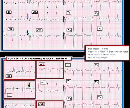
Therefore the impulse must have originated somewhere lower in the atria, perhaps near the coronary sinus. The April 17, 2022 post ( Leads V1,V2 misplacement ). The May 5, 2022 post ( LA-RA reversal ). The May 24, 2022 post ( LA-LL reversal ). The May 26, 2022 post ( LA-LL reversal ).
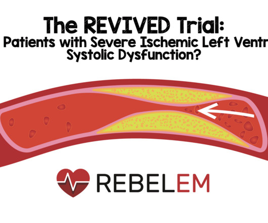
Background: Coronary artery disease can result in hibernating myocardium (chronic myocardial contractile dysfunction) due to ischemia. The theory is that there is reduced coronary blood flow and increased myocardial demand resulting in impaired contractility. Paper: Perera D et al. OMT: 38.0% HR 0.99; 95% CI 0.78 to 1.27; p = 0.96
The De Winter ECG pattern: morphology and accuracy for diagnosing acute coronary occlusion: systematic review. Hayakawa A, Tsukahara K, Miyagawa S, et al. Published 2022 Feb 20. Immediate and early percutaneous coronary intervention in very high-risk and high-risk non-ST segment elevation myocardial infarction patients.
And now this finding is even formally endorsed as a "STEMI equivalent" in the 2022 ACC guidelines!!! Association of intravenous morphine use and outcomes in acute coronary syndromes: Results from the CRUSADE Quality Improvement Initiative. de Winter et al in N Engl J Med 359:2071-2073, 2008. Am Heart J. 2005;149:1043–1049.
He underwent coronary angiography which showed severe multivessel disease, and he agreed to proceed with workup for CABG. The October 21, 2022 post — for " artifactual VT". Additional review of ECG artifacts by Pérez-Riera et al ( Ann Noninvasic Electrocardiol 23:e12494, 2018 ) VT Artifact — by Knight et al: NEJM 341:1270-1274, 1999.
Herzog et al. Khan et al. Problem #1: As I emphasized in My Comment in the December 6, 2022 post — Not all patients with acute MI report chest pain. As we've often emphasized in Dr. Smith's ECG Blog — Posterior leads often provide false reassurance ( See My Comment at the bottom of the page in the September 21, 2022 post ).
Hospital Course The patient was taken emergently to the cath lab which did not reveal any significant coronary artery disease, but she was noted to have reduced EF consistent with Takotsubo cardiomyopathy. Reference on Troponins: Xenogiannis I, Vemmou E, Nikolakopoulos I, et al. It can only be seen by IVUS. MINOCA has many etiologies.
Hgb 11g/dL (110g/L) and leukocytosis, and a mildly elevated troponin (36 ng/L, with normal 1mm STE in aVR due to ACS will require coronary artery bypass surgery for revascularization, the infarct artery is often not the LM, but rather the LAD or severe 3-vessel disease. Harhash AA, Huang JJ, Reddy S, et al. Knotts et al.
The coronary angiography showed a 100% ostial main (obtuse) marginal occlusion!" Dominant right coronary, atherosclerotic and calcified. Presence of a single coronary lesion: occlusion of the ostial main marginal.
A Short Comment on PIRP and T Waves: Oliva et al found a strong association of myocardial rupture with postinfarction regional pericarditis. Another possible cause of pseudonormalization of T waves mentioned many times on this blog is the pseudonormalization caused by re-occlusion of an infarct related reperfused coronary artery.
As I emphasized in My Comment at the bottom of the page in the October 10, 2022 post in Dr. Smith's ECG Blog — Interpretation of a post-resuscitation ECG can be extremely challenging. Many of these patients have preexisting coronary and other forms of severe heart disease. First — Some thoughts on the post -resuscitation ECG.
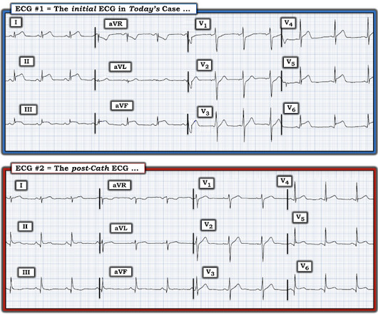
Clinical Question : In patients who suffer an OHCA without ST-segment elevation on the post-resuscitation ECG, will early coronary angiogram (CAG) vs. delayed CAG improve outcomes? Article: How-Berlemont C, Lamhaut L, Diehl J, et al. Published online June 08, 2022. JAMA Cardiol. P., & Cariou, A. PMID: 26453685 Vyas, A.,
Coronaries were normal, as was serial troponin. I sent the initial ECG of both cases without any context to my colleague Mazen El-Baba, a senior EM resident with an interest in ECG interpretation, and he responded: “RBBB with first degree AV block” for the first (ie no acute coronary occlusion), and “RBBB and superimposed OMI” for the second.
STEMI criteria is bad at differentiating between normal variant and acute coronary occlusion or reperfusion, and initial troponin levels don't differentiate between occlusive and non-occlusive MI 3. the presence of J waves from early repolarization doesn’t rule out an acute coronary occlusion 4. McLaren et al, including Meyers/Smith.
But the stuttering pain and sudden onset suggest acute coronary occlusion (Occlusion MI, or OMI). Cath lab activation by the ED and I agree with coronary angiography emergently." Result: no angiographically significant obstructive coronary artery disease. Smith and Meyers to diagnose both obvious (STEMI) and subtle OMI.
The patient proceeded to cath where all coronaries were described as normal with no evidence of any CAD, spasm, or any other abnormality. In the largest study looking at this topic by Mizusawa et al., Recently the rate of true arrhythmic events related to fevers in the classic Brugada Type 1 syndrome was explored by Michowitz et al.
Paper 1: Schmidt HJ et al. PMID: 360027567 [ Access on Read by QxMD ] Paper 2: Kjaergaard J et al. References: Schmidt HJ et al. PMID: 360027567 [ Access on Read by QxMD ] Kjaergaard J et al. A higher MAP may offer advantages due to improved cerebral perfusion pressure, however data is lacking. Liberal O2: 33.9%
Sequence of events in angina at rest: Primary reduction in coronary flow. 2022 ACC Expert Consensus Decision Pathway on the Evaluation and Disposition of Acute Chest Pain in the Emergency Department: A Report of the American College of Cardiology Solution Set Oversight Committee. 2022 Nov, 80 (20) 1925–1960. Arch Intern Med.
It was a 60yo with a history of stents to the circumflex and right coronary arteries, who presented with 9 hours of fluctuating central chest pain. This is step 4 : relying on the first troponin level to rule out acute coronary occlusion. 4] CT revealed no dissection but extensive coronary atherosclerosis. J of Emerg Med 2021.
We organize all of the trending information in your field so you don't have to. Join 5,000+ users and stay up to date on the latest articles your peers are reading.
You know about us, now we want to get to know you!
Let's personalize your content
Let's get even more personalized
We recognize your account from another site in our network, please click 'Send Email' below to continue with verifying your account and setting a password.


Let's personalize your content