Remove 2024 Remove ALS Remove Coronary
article thumbnail

Chest pain: Are these really "Nonspecific ST-T wave abnormalities", as the cardiologist interpretation states?

Dr. Smith's ECG Blog

1] But there are multiple other abnormalities that make this ECG diagnostic of Occlusion MI, localized likely to the right coronary artery: 1. Systematic review and meta-analysis of diagnostic test accuracy of ST-segment elevation for acute coronary occlusion. Int J Cardiol 2024 2. Nikus et al. Kontos et al.

STEMI 123
article thumbnail

Is OMI an ECG Diagnosis?

Dr. Smith's ECG Blog

The ECG is just a test: a Bayesian approach to acute coronary occlusion If a patient with a recent femur fracture has sudden onset of pleuritic chest pain, shortness of breath, and hemoptysis, the D-dimer doesn’t matter: the patient’s pre-test likelihood for PE is so high that they need a CT. Amsterdam et al. Alencar et al.

STEMI 121
professionals

Sign Up for our Newsletter

This site is protected by reCAPTCHA and the Google Privacy Policy and Terms of Service apply.

article thumbnail

Another deadly triage ECG missed, and the waiting patient leaves before being seen. What is this nearly pathognomonic ECG?

Dr. Smith's ECG Blog

In fact, Kosuge et al. showed that among patients with either acute coronary syndrome or acute pulmonary embolism and negative T waves in the precordial leads (V1-V4), that inverted T waves in leads III and V1 were present in only 1% of patients with acute coronary syndrome and 88% of patients with pulmonary embolism. “The

E-9-1-1 139
article thumbnail

Palpitations while awaiting CABG

Dr. Smith's ECG Blog

He underwent coronary angiography which showed severe multivessel disease, and he agreed to proceed with workup for CABG. The native QRS may be more obvious in one lead or another. == MY Comment , by K EN G RAUER, MD ( 5/30 /2024 ): == I was not taught about artifact in medical school. Take advantage of all the telemetry leads.

article thumbnail

60 year old with chest pain, STEMI negative. What should the discharge diagnosis be?

Dr. Smith's ECG Blog

First trop was 7,000ng/L (normal 25% of ‘Non-STEMI’ patients with delayed angiography have the exact same pathology of acute coronary occlusion. The new ACC expert consensus explains that: “STEMI ECG criteria on a standard 12-lead ECG alone will miss a significant minority of patients who have acute coronary occlusion. Take home 1.

STEMI 70
article thumbnail

Guidelines would (erroneously) say that this patient who was defibrillated and resuscitated does not need emergent angiography

Dr. Smith's ECG Blog

The proof of this is that only 5% of patients enrolled had acute coronary occlusion. Lemkes JS, Janssens GN, van der Hoeven NW, et al. Coronary Angiography after Cardiac Arrest without ST-Segment Elevation. We at Hennepin recently published this study Sharma et al. This study failed to do so. 5% vs. 58%!! 5% vs. 58%!!

article thumbnail

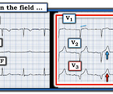
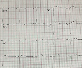
Dynamic OMI ECG. Negative trops and negative angiogram does not rule out coronary ischemia or ACS.

Dr. Smith's ECG Blog

Studies such as those by Moise et al 14 and Ellis et al 39 have shown that the relative risk of developing an acute myocardial infarction in the territory supplied by an artery with a 70%. years, with the interval as long as 12 or 18 years in some studies. Unfortunately, vascular remodeling is variable and inconsistent.