Remove 2024 Remove Coronary Remove OR
article thumbnail

Early repol or anterior OMI?

Dr. Smith's ECG Blog

Dr. Smith and other authors showed the utility of Speckle Tracking Strain Echo in this case report: Diagnosis of acute coronary occlusion in patients with non–STEMI by point-of-care echocardiography with speckle tracking Repeat ECG: Slightly less hyperacute T waves, likely indicating improving flow compared to the first ECG.

OR 112
article thumbnail

"The dye don't lie".except when it does. Angiogram Negative, or is it?

Dr. Smith's ECG Blog

You can easily imagine this patient getting one of several diagnoses -- vasospasm, MINOCA , pericarditis, or maybe even no diagnosis at all beyond "non-obstructive coronary artery disease." Another option would be to use Optical Coherence Tomography for Coronary Imaging ). That plaque is at risk of thrombosing again.

OR 70
professionals

Sign Up for our Newsletter

This site is protected by reCAPTCHA and the Google Privacy Policy and Terms of Service apply.

article thumbnail

Resuscitated from ventricular fibrillation. Should the cath lab be activated?

Dr. Smith's ECG Blog

But cardiac arrest is a period of near zero flow in the coronary arteries and causes SEVERE ischemia. or is he an ACCESS Trial Candidate? == MY Comment , by K EN G RAUER, MD ( 7/5 /2024 ): == Clinical ECG interpretation is a 2-Step process. It also does not uniformly indicate severe coronary disease.

article thumbnail

Is OMI an ECG Diagnosis?

Dr. Smith's ECG Blog

The ECG is just a test: a Bayesian approach to acute coronary occlusion If a patient with a recent femur fracture has sudden onset of pleuritic chest pain, shortness of breath, and hemoptysis, the D-dimer doesn’t matter: the patient’s pre-test likelihood for PE is so high that they need a CT. But does this matter?

STEMI 121
article thumbnail

Journal Feed Weekly Wrap-Up

EMDocs

2024 Jan;75:137-142. 2024 Jan;22(1):140-151. 3: New AI-Powered OMI Detector Spoon Feed An artificial intelligence (AI) ECG model outperformed standard STEMI criteria in identifying occlusion myocardial infarction (OMI) confirmed by coronary angiography. It’s time to learn smarter. Am J Emerg Med. doi: 10.1016/j.ajem.2023.10.049.

article thumbnail

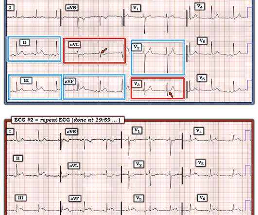
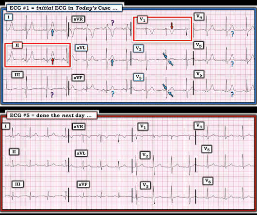
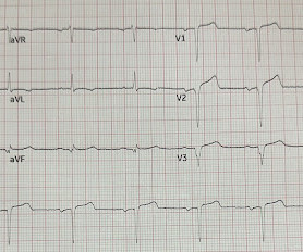
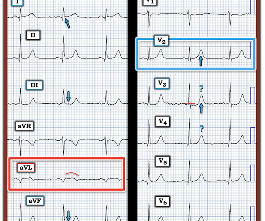
Dynamic OMI ECG. Negative trops and negative angiogram does not rule out coronary ischemia or ACS.

Dr. Smith's ECG Blog

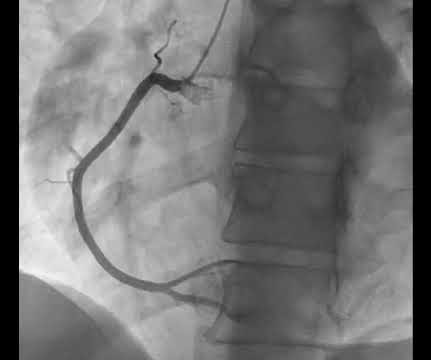
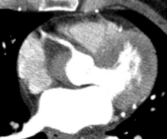
Compensatory enlargement was defined as being present when the total coronary arterial cross-sectional area at the stenotic site was greater than that at the proximal nonstenotic site. Thus, the lumen observed may actually still be the same size as the original, normal lumen. Unfortunately, vascular remodeling is variable and inconsistent.

article thumbnail

Chest pain: Are these really "Nonspecific ST-T wave abnormalities", as the cardiologist interpretation states?

Dr. Smith's ECG Blog

1] But there are multiple other abnormalities that make this ECG diagnostic of Occlusion MI, localized likely to the right coronary artery: 1. Systematic review and meta-analysis of diagnostic test accuracy of ST-segment elevation for acute coronary occlusion. Int J Cardiol 2024 2. Eur Herat J Digital Health 2024

STEMI 123