This site uses cookies to improve your experience. To help us insure we adhere to various privacy regulations, please select your country/region of residence. If you do not select a country, we will assume you are from the United States. Select your Cookie Settings or view our Privacy Policy and Terms of Use.
Cookie Settings
Cookies and similar technologies are used on this website for proper function of the website, for tracking performance analytics and for marketing purposes. We and some of our third-party providers may use cookie data for various purposes. Please review the cookie settings below and choose your preference.
Used for the proper function of the website
Used for monitoring website traffic and interactions
Cookie Settings
Cookies and similar technologies are used on this website for proper function of the website, for tracking performance analytics and for marketing purposes. We and some of our third-party providers may use cookie data for various purposes. Please review the cookie settings below and choose your preference.
Strictly Necessary: Used for the proper function of the website
Performance/Analytics: Used for monitoring website traffic and interactions
In ACS, chest pain is the warning sign of ongoing ischemia. In this case, you should get a second defibrillator and perform double sequential external defibrillation (DSED). Simply attach a second defibrillator as shown in the diagram below and deliver max shocks from both devices simultaneously.
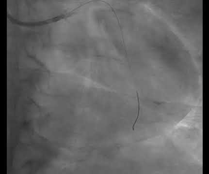
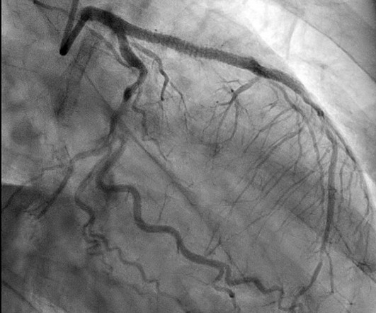
After ruling out for ACS, the patient underwent angiography where he was found to have severe stable disease, which was already known. NOTE: Although this case study is from 1995 the misdiagnosis of wide tachycardias "because the patient is stable" remains all-too-common in 2025.
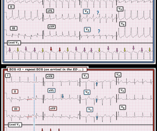
He had several older ECGs on file, here are two examples: 6 days prior: 2 months prior: In the context of ACS symptoms, and when able to compare the new vs. old ECG, the top ECG is DIAGNOSTIC of OMI until proven otherwise. Defibrillation was performed, and ROSC was achieved. Today's initial ECG is not an easy tracing to interpret.
Whenever I see PVCs with the morphology and axis seen in todays case I always look for signs of AC ( Arrhythmogenic Cardiomyopathy ). See this case for an in-depth discussion of AC and an example of VT and ECG changes associated with this disorder. Arrhythmogenic cardiomyopathy often manifests with PVCs from the RV. 3] Lavalle, C.
Several 200 J shocks did not terminate the VF, so a second defibrillator was applied for double sequential defibrillation with 400 J. She was defibrillated perhaps 25 times. After completing the ACS algorithm with amiodarone and lidocaine, there are diminishing returns on further treatments. SanzRuiz, R., Solis, J., &
We organize all of the trending information in your field so you don't have to. Join 5,000+ users and stay up to date on the latest articles your peers are reading.
You know about us, now we want to get to know you!
Let's personalize your content
Let's get even more personalized
We recognize your account from another site in our network, please click 'Send Email' below to continue with verifying your account and setting a password.


Let's personalize your content