This site uses cookies to improve your experience. To help us insure we adhere to various privacy regulations, please select your country/region of residence. If you do not select a country, we will assume you are from the United States. Select your Cookie Settings or view our Privacy Policy and Terms of Use.
Cookie Settings
Cookies and similar technologies are used on this website for proper function of the website, for tracking performance analytics and for marketing purposes. We and some of our third-party providers may use cookie data for various purposes. Please review the cookie settings below and choose your preference.
Used for the proper function of the website
Used for monitoring website traffic and interactions
Cookie Settings
Cookies and similar technologies are used on this website for proper function of the website, for tracking performance analytics and for marketing purposes. We and some of our third-party providers may use cookie data for various purposes. Please review the cookie settings below and choose your preference.
Strictly Necessary: Used for the proper function of the website
Performance/Analytics: Used for monitoring website traffic and interactions
Encourage your ED to set up an algorithm that you can follow based on your laboratory’s assay. Low-risk patients do not routinely require stress testing in the ED. You (or someone in your department) needs to know which assay your ED has, and use the appropriate values for that assay. Otherwise, apply a simplified approach.
Trauma season is at hand and like all other pediatric emergency departments in the country, we find our ED breaking ( pun intended ) at the seams with orthopedic injuries. Davidson JS, Brown DJ, Barnes SN, et al. West S, Andrews J, Bebbington A, et al. Symons S, Rowsell M, Bhowal B, et al. J Pediatr Orthop. 2008;24:65–70.
Date: September 23, 2024 Reference: Essat et al. Case: You are working a busy shift in a rural emergency department (ED) and your excellent Family Medicine trainee presents a case of a 63-year-old woman with chest pain and some intermittent radiation into the inter-scapular region. Reference: Essat et al.
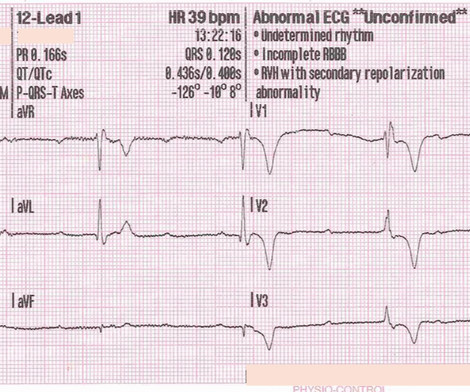
male presents to the ED at 6:45 AM with left sided chest dull pressure that woke him up from sleep at 3am. He arrived to the ED at around 6:45am, and stated the pain has persisted. Here is his ED ECG at triage: Obvious high lateral OMI that does not quite meet STEMI criteria. The pain radiated to both shoulders.
100% seems too good to be true Morello et al., European Journal of Internal Medicine , [link] You can listen to my 27-minute rant on Youtube here: [link] This multinational trial looked at a three-pronged diagnostic protocol in the ED for adults with suspected acute aortic syndromes. Did they pick a heap of PEs? Emerg Med J.
You turn to the attending and ask, “do you really think this could be acute coronary syndrome (ACS)?” ACS is usually amongst this differential, as cardiovascular disease is a leading cause of morbidity and mortality in this population. Reference: Wang et al. The utility of troponin testing to diagnose or exclude ACS. *
What Your Gut Says: The patient has a tachydysrhythmia which may be the presentation of acute coronary syndrome (ACS) even though the patient has no ischemic symptoms. Type 2 MI is common in the ED and can result from vigorous exercise (common in athletes after marathons), sepsis, trauma and tachydysrhythmias including SVT.
Date: September 8th, 2021 Reference: Desch et al. Date: September 8th, 2021 Reference: Desch et al. He is interested and experienced in healthcare informatics, previously worked with ED-directed EMR design, and is involved in the New York City Health and Hospitals Healthcare Administration Scholars Program (HASP).
Reference: Brichko et al. Reference: Brichko et al. Case: A 46-year-old female presents to the emergency department (ED) with sudden onset, severe right flank pain. Delays in providing adequate analgesia leads to poorer patient outcomes, prolonged ED length of stay and reduced patient satisfaction (17, 18). AEM Feb 2021.
Other causes of sickling: acidosis, dehydration, inflammation, infection, fever, and blood stasis Sickling leads to vascular occlusion, end-organ ischemia, and decreased RBC lifespan, which, in turn, leads to pain crisis, acute anemia, sequestration, infection, and acute chest syndrome (ACS.) Each episode of ACS has a 9% mortality rate.
In fact, Kosuge et al. Stein et al. This is a paper worth reading : Marchik et al. Kosuge et al. showed that , when T-waves are inverted in precordial leads, if they are also inverted in lead III and V1, then pulmonary embolism is far more likely than ACS. Witting et al. of patients with PE and 3.3%
Although the attending crews did not consider the ECG pathognomonic for occlusive thrombosis, they nonetheless considered the patient high-risk for ACS and implored him to reconsider. Here is the final ECG just prior to ED transfer. It’s important to stress the presence of a normal QRS (i.e., The pathology is now painfully evident.
Date: June 30th, 2022 Reference: McGinnis et al. Date: June 30th, 2022 Reference: McGinnis et al. Case: You are working a shift in your local community emergency department (ED) when a 47-year-old male presents with chest pain. Background: Chest pain is one of the most common presentations to the ED. AEM June 2022.
Sickling leads to vascular occlusion, end-organ ischemia, and decreased RBC lifespan, which, in turn, leads to pain crisis, acute anemia, sequestration, infection, and acute chest syndrome (ACS). ACS is lung injury due to vaso-occlusion in the pulmonary vasculature; many with ACS will have a concomitant vaso-occlusive pain crisis.
However, RSI has never been shown to reduce the risk of aspiration in the ED (13) or during emergent OR cases (14). While RSI should remain the gold standard in the vast majority of patients in the ED, FI presents an additional technique to mitigate anatomic or physiologic risk. To date, ketamine has been the agent of choice (12).
It should be emphasized here that this is a presentation of high-pretest probability for Acute Coronary Syndrome (ACS). ACS and hyperkalemia both have lethal downstream consequences, so it is imperative for the clinician to acclimate to the presentation, or developing, features of each. link] [1] Zachary et al. 2] Costanzo, L.
Reference: Jhunjhunwala et al. Reference: Jhunjhunwala et al. Upon arrival in the emergency department (ED), the patient is incoherently speaking, has a pulse of 135 beats per minute, blood pressure of 85/50 mm Hg, and an obvious open wound in their left mid-axillary line at the level of the nipple.
Notoriously elusive, with a high misdiagnosis rate, thoracic aortic dissection (AD) can mimic many conditions, including acute coronary syndrome (ACS, the most common), gastroesophageal reflux disease (GERD), stroke, and spinal-cord compression. The patient is admitted for ACS to a cardiologist who says he will see the patient in the morning.
Date: May 24th, 2022 Reference: Broder et al. Date: May 24th, 2022 Reference: Broder et al. The writing group of GRACE-2 wanted to look at clinically relevant questions to address the care of adult patients with low-risk, recurrent, previously undifferentiated abdominal pain in the ED. Reference: Broder et al.
[link] Case continued She arrived in the ED and here is the first ED ECG. Angiogram No obstructive epicardial coronary artery disease Cannot exclude non-ACS causes of troponin elevation including coronary vasospasm, stress cardiomyopathy, microvascular disease, etc. Lindahl et al. From Gue at al.
1] Here is the admitting ED ECG after cancellation of Code STEMI. The patient continued to verbalize cessation of symptoms while in the ED. However, in this context (i.e. cessation of symptoms), and in this unique territory (V2-V4), the T-wave characteristics are consistent with posterior reperfusion! [1] 1] Driver, B. 3] Niu, T.,
He advises, however, recurrent syncopal episodes for the past six months, some of which have resulted in ED admission, yet no identifying mechanism could be determined. Chou’s Electrocardiography in Clinical Practice (6th ed). Josephson’s Clinical Cardiac Electrophysiology: Techniques and Interpretations (6th ed). 2] Meyers, H.
The ECG’s were sent to the PCI center, and the providers in the respective ED identified the T wave characteristics mentioned above. Such aggressive investigation was particularly warranted in this case because of symptoms compatible with ACS, as well as an equally frightening revelation of family history. link] [1] Mirand, D.
Episode 108: Unexplained Sinus Tachycardia Mental Model Background: When a patient in the ED has sinus tachycardia our job as emergency physicians is to identify and treat of the underlying pathology. Lactate Troponin Could this be ACS or myocarditis? In the ED, our job is to identify and treat underlying causes. Ann Emerg Med.
There were zero patients in this study with a "normal" ECG who had any kind of ACS! Deutch et al. Figure-1: I've labeled the initial ECG in the ED. KEY Point: All patients who present to the ED for new CP should promptly have a triage ECG recorded, that is then immediately interpreted by the ED physician.
The Queen of Hearts agrees: Here the Queen explains why: However, it was not interpreted correctly by the providers: ED interpretation of ECG: "paced rhythm, LBBB but no STEMI pattern." The cath report showed: Significant stenosis with subtotal occlusion (99%) in the prox to mid Lcx, culprit of ACS, TIMI flow 1. 2021;23:187.
Antonaci L, et al. Tritos NA, et al. Levi M, et al. Fishbein MH, et al. Cetinkaya PG, et al. Niu T, et al. Verkuijl SJ, et al. Varni JW, et al. Dias FC, et al. Peter C, et al. Ahlberg R, et al. Shir A, et al. Kuypers KLAM, et al. Hegeman EM, et al.
Cardiology consult note written around that time documents that "Pain improved with NTG, morphine in ED but still present." The original term " benign early repolarization" has fallen out of favor since the seminal paper by Haïssaguerre et al. We therefore need to assume and rule "out" ACS — more than having to rule it "in".
Key: Consider eclampsia in any pregnant and postpartum woman presenting to the ED, especially in patients with symptoms such as headache, confusion/altered mental status, vision changes, and hypertension. ED Evaluation: Assessment focuses on looking for complications and mimics. Wilkerson RG, Ogunbodede AC. 2019;37(2):301-316.
Anecdotally, had there been symptoms unequivocally consistent with ACS then one could justifiably make the case for a potential D1 occlusion. Chou’s Electrocardiography in Clinical Practice, 6th ed. 4] Baranchuk, A, et al. Goldberger’s Clinical Electrocardiography: A Simplified Approach, 9th ed. 2] Surawicz, B.
His parent noticed a dental problem and immediately brought the patient to the ED. The patient was walking his dog when it ran after a squirrel. The patient was pulled forward, causing him to hit his tooth on the asphalt. The physical exam is shown below. Which of the following is the best next recommendation? Dent Traumatol.
A man in his 70s with past medical history of hypertension, dyslipidemia, CAD s/p left circumflex stent 2 years prior presented to the ED with worsening intermittent exertional chest pain relieved by rest. In our opinion it should not be given in ACS unless you are committed to the cath lab. Hayakawa A, Tsukahara K, Miyagawa S, et al.
Written by Jesse McLaren A 75 year-old patient with diabetes and end stage renal disease was sent to the ED after dialysis for three days of nausea, vomiting, loose stool, lightheadedness and fatigue. But because the patient had no chest pain or shortness of breath, it was not deemed to be from ACS. Herzog et al. Khan et al.
The NIHSS cutoff that predicts outcomes is 4 points higher in AC compared with PC infarctions. Median time from ED arrival to diagnosis was 8 hours 24 min in one study, with only 19% being diagnosed within the 4.5-hour References: Gaillard F, Glick Y, Tatco V, et al. Post TW, ed. 61.4.496 Navi BB, Kamel H, Shah MP, et al.
Because there was proven thrombus (ACS) but the troponin never went above the 99% reference range (and therefore cannot be called MI -- definition of MI requires rise and/or fall of troponin with at least one value above the 99% reference range), this is UNSTABLE ANGINA with ST Elevation. After all, there is no S wave in lead V3.
ECG 1 at time zero EARLY REPOLARIZATION ABNORMAL ECG ED final official overread: "early repol vs hyperacute T, minimal changes from previous (previous shown below)" What do YOU think? Lemkes et al. No wall motion abnormality This shows that significant ACS can have ZERO WMA!! A 70-something y.o. Eur Heart J 2018. Full text link.
Smith: If this is ACS (a big if), t his is just the time when one should NOT use "upstream" dual anti-platelet therapy ("upstream" means in the ED before angiography). History sounds concerning for ACS (could be critical stenosis, triple vessel), but differential also includes dissection, GI bleed, etc. Knotts et al.
Smith and Meyers answer: First , LM occlusion is uncommon in the ED because most of these die before they can get a 12-lead recorded. She had a proven 100% Left Main occlusion No ST shift in aVR This pattern of RBBB/LAFB was also the most common pattern in Fiol et al. Widimsky P et al. Knotts et al. TIMI 0/1 flow).(61,62)
ED treatment should focus on airway, breathing, and circulation with consideration for cervical spine protection depending on the circumstances surrounding the event. References Webb AC, Wheeler A, Ricci A, et al. Pellegrino F, Raffaldi I, Rossi R, et al. Chotai PN, Manning L, Eithun B, et al. South Med J.
link] A 30 year-old woman was brought to the ED with chest pain. SCAD isn’t rare, especially in women Historically SCAD had been identified in 22% of ACS cases in women. Pregnancy is not a common cause of SCAD When ACS occurs in the peripartum period, SCAD is responsible in 43% of cases. A study by Hassan et al.
All you know, back in ED, is that the ETA is 10 minutes, and there is a single stab wound to the chest. The ODP is caught up leaving theatres and has not yet made it down to ED. Back in ED with Ranulf, and pack two has gone through. Laan DV, Vu TD, Thiels CA et al. The trauma call goes out. Trauma Surg Acute Care Open.
She arrived in the ED 37 minutes after 911 was called, with continuing CPR. Here is an article I wrote: Updates on the ECG in ACS. Was this: 1) ACS with ischemia and spontaneous reperfusion? Fine ventricular fibrillation She received 2 mg epinephrine, 150 mg amiodarone and underwent chest compressions with the LUCAS device.
A 44 year-old male with unknown past medical history came by emergency medical services (EMS) to the emergency department (ED) for an electrical injury and fall from a high voltage electrical pole. 2,3,5 Except for laundry or electrical car outlets (240 V AC), all U.S. household outlets are rated at 120 V AC. 2023 Jul 17.
We organize all of the trending information in your field so you don't have to. Join 5,000+ users and stay up to date on the latest articles your peers are reading.
You know about us, now we want to get to know you!
Let's personalize your content
Let's get even more personalized
We recognize your account from another site in our network, please click 'Send Email' below to continue with verifying your account and setting a password.


Let's personalize your content