This site uses cookies to improve your experience. To help us insure we adhere to various privacy regulations, please select your country/region of residence. If you do not select a country, we will assume you are from the United States. Select your Cookie Settings or view our Privacy Policy and Terms of Use.
Cookie Settings
Cookies and similar technologies are used on this website for proper function of the website, for tracking performance analytics and for marketing purposes. We and some of our third-party providers may use cookie data for various purposes. Please review the cookie settings below and choose your preference.
Used for the proper function of the website
Used for monitoring website traffic and interactions
Cookie Settings
Cookies and similar technologies are used on this website for proper function of the website, for tracking performance analytics and for marketing purposes. We and some of our third-party providers may use cookie data for various purposes. Please review the cookie settings below and choose your preference.
Strictly Necessary: Used for the proper function of the website
Performance/Analytics: Used for monitoring website traffic and interactions
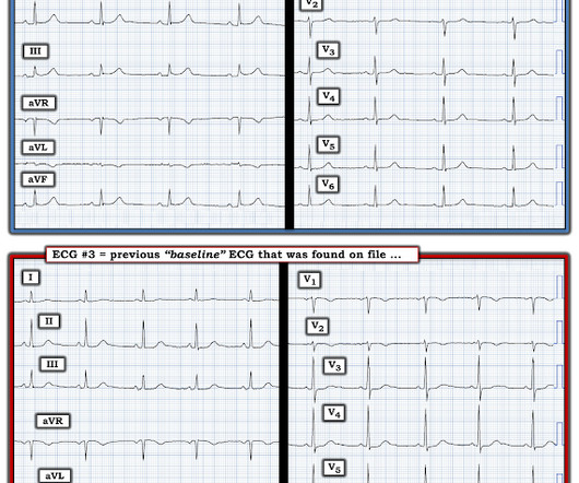
Figure 1-1 My colleague, a faithful student of ECG interpretation, handed me the tracing and said that it warranted STEMI activation because of apparent terminal QRS distortion (TQRSD) in V2. Anecdotally, had there been symptoms unequivocally consistent with ACS then one could justifiably make the case for a potential D1 occlusion.
Smith : there is some minimal ST elevation in V2-V6, but does not meet STEMI criteria. Transient STEMI has been studied and many of these patients will re-occlude in the middle of the night. No wall motion abnormality This shows that significant ACS can have ZERO WMA!! Is it normal STE? This is a "Transient OMI".
Note 2 other similar cases at the bottom that come from my book, The ECG in Acute MI. This meets "STEMI criteria" However, there is very high voltage, with a very deep S-wave in V2 and tall R-wave in V4. The morphology is not right for STEMI. This is very good evidence that the ST elevation is not due to STEMI.
Troponin T peaked at 2074 ng/L (very high, typical of OMI/STEMI). Here is an example of isolated RV infarction, from Dr. Smith's book : Learning points: 1) OMI can be very subtle and RV infarction may manifest poorly on the standard ECG. Post PCI the patient became gravely hypotensive and "shocky". The LV EF was 57% at formal echo.
Discussion: This case highlights many important points worthy of discussion, mainly because it represents very routine care for ACS but there are so many ways we could improve outcomes with tools we already have! Limitations of registry data: This patient presented with STEMI (-) OMI and developed STEMI the following day.
We organize all of the trending information in your field so you don't have to. Join 5,000+ users and stay up to date on the latest articles your peers are reading.
You know about us, now we want to get to know you!
Let's personalize your content
Let's get even more personalized
We recognize your account from another site in our network, please click 'Send Email' below to continue with verifying your account and setting a password.


Let's personalize your content