This site uses cookies to improve your experience. To help us insure we adhere to various privacy regulations, please select your country/region of residence. If you do not select a country, we will assume you are from the United States. Select your Cookie Settings or view our Privacy Policy and Terms of Use.
Cookie Settings
Cookies and similar technologies are used on this website for proper function of the website, for tracking performance analytics and for marketing purposes. We and some of our third-party providers may use cookie data for various purposes. Please review the cookie settings below and choose your preference.
Used for the proper function of the website
Used for monitoring website traffic and interactions
Cookie Settings
Cookies and similar technologies are used on this website for proper function of the website, for tracking performance analytics and for marketing purposes. We and some of our third-party providers may use cookie data for various purposes. Please review the cookie settings below and choose your preference.
Strictly Necessary: Used for the proper function of the website
Performance/Analytics: Used for monitoring website traffic and interactions
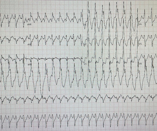
David Didlake @DidlakeDW EMS personnel responded to the residence of an 81 y/o Male with syncope. 2. Coronary angiography reveals significant and severe CAD involving all three epicardial vessels. Access the links provided for a detailed review of varying ECG patterns when ACS breaches the typical subendocardial ischemia pattern.
He denied any known history of CAD, but did report ASCVD risk factors to include HTN, HLD, and DM. I interpreted the ECG as VT with two primary etiological possibilities: 1. Abrupt plaque ulceration of Type 1 ACS leading to VT.
Moreover, he had no pertinent medical history to report in terms of CAD, HTN, HLD, or DM, for example. According to the EMS narrative, this patient initially refused hospital transport and advised that he would seek evaluation at a later time with his personal physician. A 12 Lead ECG was recorded. A 12 Lead ECG was recorded.
A 63 year old man with a history of hypertension, hyperlipidemia, prediabetes, and a family history of CAD developed chest pain, shortness of breath, and diaphoresis after consuming a large meal at noon. He called EMS, who arrived on scene about two hours after the onset of pain to find him hypertensive at 220 systolic.
Category 1 : Sudden narrowing of a coronary artery due to ACS (plaque rupture with thrombosis and/or downstream showering of platelet-fibrin aggregates. Smith : This is ACS even if the troponin returns normal, and the first troponin especially might return normal. This results in Type I MI. She was urgently taken to the Cath Lab.
Sent by Anonymous, written by Pendell Meyers A man in his 60s with history of CAD and 2 prior stents presented to the ED complaining of acute heavy substernal chest pain that began while eating breakfast about an hour ago, and had been persistent since then, despite EMS administering aspirin and nitroglycerin. Pre-intervention.
A man in his 70s with past medical history of hypertension, dyslipidemia, CAD s/p left circumflex stent 2 years prior presented to the ED with worsening intermittent exertional chest pain relieved by rest. In our opinion it should not be given in ACS unless you are committed to the cath lab. He was diagnosed as NSTEMI.
Similarly, if a patient with known CAD presents with refractory ischemic chest pain, the ECG barely matters: the pre-test likelihood of acute coronary occlusion is so high that they need an emergent angiogram. 1] European guidelines add "regardless of biomarkers".
This case was provided by Spencer Schwartz, an outstanding paramedic at Hennepin EMS who is on Hennepin EMS's specialized "P3" team, a team that receives extra training in advanced procedures such as RSI, thoracostomy, vasopressors, and prehospital ultrasound. I could have told you this (and did tell you this) without an MRI.
It was edited by Smith CASE : A 52-year-old male with a past medical history of hypertension and COPD summoned EMS with complaints of chest pain, weakness and nausea. En route, EMS administered aspirin 325mg by mouth, but withheld nitroglycerin due to initial hypotension. Answer below in the still shot.
EMS found the patient in VFib and performed ACLS for 26 minutes then obtained ROSC. The patient was transferred immediately for angiogram which revealed no significant CAD, and no intervention was performed. Learning Points: The myocardium doesn't know the etiology of OMI (ACS, spasm, dissection, embolus, etc.),
He reported to EMS a medical history of GERD only. Furthermore, there was no family history of early CAD, MI, or sudden cardiac death. V2 – in the final EMS ECG the ST segment was baseline. V3 – in the final EMS ECG the ST segment was still slightly depressed. However, in this context (i.e.
If this is ACS with Aslanger's pattern , the ST depression vector of subendocardial ischemia (due to simultaneous 3 vessel or left main ACS) is directed toward lead II (inferior and lateral). Thus, this apparently is Aslanger's Pattern (inferior OMI with single lead STE in lead III, with simultaneous subendocardial ischemia).
A middle-aged male with h/o CAD and stents presented with typical chest pressure. EMS recorded the following ECG: What do you see? It is highly associated with proximal LAD occlusion or severe left main ACS and with bad outcomes. This is a very common misread. It may be difficult to read STEMI in the setting of RBBB.
She did not receive any opioids (which would mask her pain without affecting any underlying ACS). She also had non-acute CAD of the left main (50%) and LCX (75%). J of National Association of EMS Physicians 2014. She was asymptomatic at the time of this ECG recorded on arrival to our ED: What do you think? They opened it.
We organize all of the trending information in your field so you don't have to. Join 5,000+ users and stay up to date on the latest articles your peers are reading.
You know about us, now we want to get to know you!
Let's personalize your content
Let's get even more personalized
We recognize your account from another site in our network, please click 'Send Email' below to continue with verifying your account and setting a password.


Let's personalize your content