This site uses cookies to improve your experience. To help us insure we adhere to various privacy regulations, please select your country/region of residence. If you do not select a country, we will assume you are from the United States. Select your Cookie Settings or view our Privacy Policy and Terms of Use.
Cookie Settings
Cookies and similar technologies are used on this website for proper function of the website, for tracking performance analytics and for marketing purposes. We and some of our third-party providers may use cookie data for various purposes. Please review the cookie settings below and choose your preference.
Used for the proper function of the website
Used for monitoring website traffic and interactions
Cookie Settings
Cookies and similar technologies are used on this website for proper function of the website, for tracking performance analytics and for marketing purposes. We and some of our third-party providers may use cookie data for various purposes. Please review the cookie settings below and choose your preference.
Strictly Necessary: Used for the proper function of the website
Performance/Analytics: Used for monitoring website traffic and interactions
Ischemia from ACS causing the chest discomfort, with VT another consequence (or coincidence)? Cardioversion will address the rhythm problem immediately, also if the chest discomfort subsides when SR is restored, ischemia from ACS becomes much less likely. In either case, prompt cardioversion is indicated.
Early coronary angiography in post-CA patients with no ST-segment elevation on the presenting ECG may still be of benefit by potentially salvaging myocardium and decreasing the incidence of systolic heart failure in survivors (95.7%, 22/23). Digestive Management Takeaway: Start enteral feeds when the patient gets to the ICU.
Immediate and early percutaneous coronary intervention in very high-risk and high-risk Non-STEMI patients. Smith comment: We have shown that use of opiates is associated with worse outcomes in ACS: Bracey, A. Opioids in ACS may reduce the pain score, but do not provide reperfusion for ongoing ACS. Lupu L, et al.
Wellens pattern is a term which refers to coronary reperfusion morphology in the anterior leads) The best answer is because the entire gestalt of the ECG shows acute right heart strain instead, and just does not look like Wellens after you've seen Wellens hundreds of times. looked at consecutive patients with PE, ACS, or neither.
A 68-year-old male with a past medical history of hypertension, diabetes mellitus, and coronary artery disease with a drug eluting stent placed 2 months ago presents with dizziness and vomiting that began 3 hours ago. The NIHSS cutoff that predicts outcomes is 4 points higher in AC compared with PC infarctions.
Written by Pendell Meyers A man in his late 40s with several ACS risk factors presented with a chief complaint of chest pain. The cardiologists felt that the ECG did not represent ACS, and thought it was more likely pericarditis, so they did not take him to the cath lab. in the ICU but survived with excellent function.
If she had no risk factors, it is doubtful that she would have developed such extensive coronary artery disease as we see on the angiogram. I took part in her ICU care and she was extubated and stable to transfer to a stepdown unit after a few days. Her repeat ECHO showed an improving EF of 37%.
The patient was upgraded to the ICU for closer monitoring. Electrocardiographic Differentiation Between Acute Pulmonary Embolism and Acute Coronary Syndromes on the Basis of Negative T Waves - ScienceDirect. looked at consecutive patients with PE, ACS, or neither. In fact, Kosuge et al. In fact, Kosuge et al. Kosuge et al.
Moreover, the Queen is only supposed to be used with a high pretest probability of ACS/OMI. The patient was admitted to the ICU for close monitoring and electrolyte repletion and had an uneventful hospital course. Instead — it commonly reflects ischemia from severe underlying coronary disease. Magnesium later resulted at 0.8
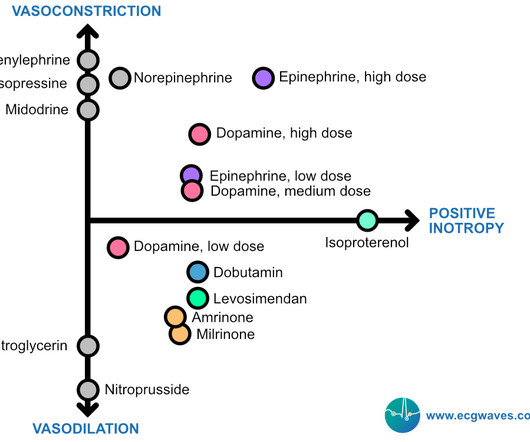
Below follows a drug manual for use in the CCU (coronary care unit), ICU (intensive care unit) or ER (emergency room). μg/kg/min + + + ++ Low dose dopamine stimulates D1 receptors and induces vasodilation in coronary, renal, cerebral and mesenteric vessels. Increases coronary blood flow. Coronary flow enhanced.
She did not receive any opioids (which would mask her pain without affecting any underlying ACS). If for some reason the angiogram is delayed, they should receive maximal medical therapy in an ICU setting with continuous 12-lead ST segment monitoring under the close attention of a practitioner with advanced ECG interpretation training.
3,10 Coronary Allograft Vasculopathy Nicknamed “The Achilles Heel of Heart Transplantation,” this accounts for the majority of patient mortality in the 5-10 year range. 10 It affects the whole length of the vessel and all layers of the coronary vasculature rather than just the intima, which is seen in non-transplant atherosclerosis.
. __ Thus , in a patient presenting with symptoms of ACS, this EKG is diagnostic of subacute LAD occlusion, possibly reperfused. Does this patient have ACS Symptoms? The patient was transferred to the ICU on pressors, where a repeat bedside echo showed an LVEF of 10-15%. The blue arrow shows a 90% stenosis of the proximal RCA.
We organize all of the trending information in your field so you don't have to. Join 5,000+ users and stay up to date on the latest articles your peers are reading.
You know about us, now we want to get to know you!
Let's personalize your content
Let's get even more personalized
We recognize your account from another site in our network, please click 'Send Email' below to continue with verifying your account and setting a password.


Let's personalize your content