This site uses cookies to improve your experience. To help us insure we adhere to various privacy regulations, please select your country/region of residence. If you do not select a country, we will assume you are from the United States. Select your Cookie Settings or view our Privacy Policy and Terms of Use.
Cookie Settings
Cookies and similar technologies are used on this website for proper function of the website, for tracking performance analytics and for marketing purposes. We and some of our third-party providers may use cookie data for various purposes. Please review the cookie settings below and choose your preference.
Used for the proper function of the website
Used for monitoring website traffic and interactions
Cookie Settings
Cookies and similar technologies are used on this website for proper function of the website, for tracking performance analytics and for marketing purposes. We and some of our third-party providers may use cookie data for various purposes. Please review the cookie settings below and choose your preference.
Strictly Necessary: Used for the proper function of the website
Performance/Analytics: Used for monitoring website traffic and interactions
He was defibrillated into VT. He then underwent dual sequential defibrillation into asystole. Then assume there is ACS. See these related cases: Cardiac arrest, defibrillated, diffuse ST depression and ST Elevation in aVR. This patient was witnessed by bystanders to collapse. They started CPR. sodium bicarbonate.
Multiple attempts at defibrillation, epinephrine, and amiodarone have been unsuccessful. Problem What is the best defibrillation strategy to treat refractory ventricular fibrillation? 2,3 Multiple published studies have addressed treatment of ventricular fibrillation with defibrillation and medications such as amiodarone and lidocaine.
Does this patient have ACS? Again, it is common to have an ECG that shows apparent subendocardial ischemia after resuscitation from cardiac arrest, after defibrillation, and after cardioversion. He did not have ACS. The remainder were due to other etiologies, (including NonSTEMI ACS). Learning Points: 1.
He was defibrillated, but they also noticed that he was being internally defibrillated and then found that he had an implantable ICD. He was unidentified and there were no records available After 7 shocks, he was successfully defibrillated and brought to the ED. There was no bystander CPR. The QRS is extremely wide.
He was resuscitated with chest compressions and defibrillation and 1 mg of epinephrine. ACS would be highly unusual in a young athlete, and given the information on his race bib, one must first suspect that the abnormal ST elevation is due to demand ischemia, not ACS. On his bib it stated that he had a congenital heart disorder.
Defibrillation is the treatment of choice in these cases but does not often result in sustained ROSC ( Kudenchuk et al 2006). Acute coronary syndrome (ACS) is responsible for the majority (60%) of all OHCAs in patients. Half of these arrests are witnessed with the other half being un-witnessed.
With respect to timing, for cardiac arrest with a shockable rhythm, it may be reasonable to administer epinephrine after initial defibrillation attempts have failed. Consider administering epinephrine after defibrillation in those with shockable rhythms. COR 2b, LOE C-LD. COR 3, No benefit, LOE B-R. COR 2b, LOE B-R. COR 2b, LOE C-LD.
She was found to be in ventricular fibrillation and was defibrillated 8 times without a single, even transient, conversion out of fibrillation. She was immediately intubated during continued compressions, then underwent a 9th defibrillation, which resulted in an organized rhythm at 42 minutes after initial arrest. References : 1.
But because Dr. Mastoras recognized the hyperacute T waves, the patient was immediately seen, the polymorphic VT was immediately defibrillated, and the patient was rapidly diagnosed and treated. The risk of SCAD is even higher in pregnancy — accounting for over 40% of angiograms performed for ACS during the peripartum period.
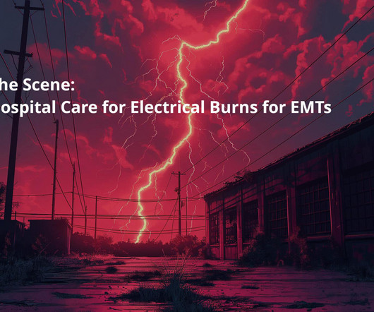
Assessing the Severity The severity of an electrical burn depends on several factors: the type of current (AC or DC), voltage, the pathway of the current through the body, the duration of contact, and the victim’s overall health. As EMTs, we’re always prepared to address these life-threatening complications alongside the burns.
The fire department, who operate at an EMT level in this municipality, arrived before us and administered 324 mg of baby aspirin to the patient due to concern for ACS. She was defibrillated and resuscitated. Most studies examine undifferentiated ACS cohorts, with only a handful providing separate data. References: 1.
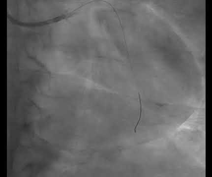
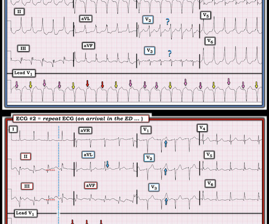
He was found in ventricular fibrillation and defibrillated, then brought to a local ED which does not have a cath lab. He was treated medically for ACS and did not get an angiogram within 72 hours. The patient was defibrillated, and then taken to the nearest ED where ECG #1 was obtained ( Figure-1 ).
Recall that, in the setting of ACS symptoms, ST depression that are maximal in leads V1-V4 (as opposed to V5 and V6) not attributable to an abnormal QRS complex is specific for OMI. When the ICD was finally interrogated, the syncopal events and shocks correlated with two VF events that were defibrillated successfully.
12 minutes later, the patient went back into VFib arrest and underwent another 15 minutes of resuscitation followed by successful defibrillation and sustained ROSC. In total, he received approximately 40 minutes of CPR and 7 defibrillation attempts. That said, ACS is by far the most common and treatable cause.
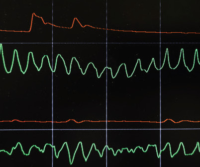
The ST segment changes are compatible with severe subendocardial ischemia which can be caused by type I MI from ACS or potentially from type II MI (non-obstructive coronary artery disease with supply/demand mismatch). The arrhythmia spontaneously converted before defibrillation was achieved.
This is diagnostic of ACS; it appears to be a reperfused acute inferior OMI. VF was refractory to amiodarone, lidocaine, double-sequential defibrillation, esmolol, etc. Then the patient would have been taken to the critical care area with a defibrillator at his side while waiting for the cath lab to be ready.
It was reportedly a PEA arrest; there was no recorded V Fib and no defibrillation. If this is ACS with Aslanger's pattern , the ST depression vector of subendocardial ischemia (due to simultaneous 3 vessel or left main ACS) is directed toward lead II (inferior and lateral). CPR was initiated immediately.
During angiogram in the cath lab, the patient suffered two episodes of ventricular fibrillation for which he was successfully defibrillated. ST depression maximal in V1-V4, without a QRS abnormality clearly causing it, in the setting of ACS symptoms, is very concerning for posterior MI until proven otherwise.
She was never seen to be in ventricular fibrillation and was never defibrillated. Medics found her apneic and pulseless, began CPR, and she was found to be in asystole. With ventilations and epinephrine, she regained a pulse. She was hypotensive in the ED and her bedside echo showed a normal RV and LV. BP gradually rose.
One must always be careful when looking for "baseline" ECGs, because the prior ECG on file may have been during another ACS event, as this one clearly was. He was defibrillated immediately and had return of normal mental status. Cath lab activation was cancelled but the transfer was accepted for urgent cardiology evaluation.
I B ECG monitoring should start immediately and a defibrillator must be ready. I C Glucose-lowering therapy should be considered in ACS patients with glucose levels >10 mmol/L (>180 mg/dL), while episodes of hypoglycaemia (defined as glucose levels <_3.9 STEMI , ST-segment elevation acute myocardial infarction ).
In ACS, chest pain is the warning sign of ongoing ischemia. In this case, you should get a second defibrillator and perform double sequential external defibrillation (DSED). Simply attach a second defibrillator as shown in the diagram below and deliver max shocks from both devices simultaneously.
After ruling out for ACS, the patient underwent angiography where he was found to have severe stable disease, which was already known. This would be approximately 95% of the patient's maximum predicted sinus rate. This demands an explanation -- sepsis, hemorrhage, withdrawal, etc. Calling sinus tachycardia raises more questions than answers.
He had several older ECGs on file, here are two examples: 6 days prior: 2 months prior: In the context of ACS symptoms, and when able to compare the new vs. old ECG, the top ECG is DIAGNOSTIC of OMI until proven otherwise. Defibrillation was performed, and ROSC was achieved.
But thankfully, when the clinical context is clearly and highly concerning for ongoing ischemia from ACS, this distinction doesn't matter much. Soon after the witnessed occlusion, the patient suffered ventricular fibrillation arrest, from which he was immediately resuscitated with 1 defibrillation.
Several 200 J shocks did not terminate the VF, so a second defibrillator was applied for double sequential defibrillation with 400 J. She was defibrillated perhaps 25 times. After completing the ACS algorithm with amiodarone and lidocaine, there are diminishing returns on further treatments. SanzRuiz, R., Solis, J., &
Whenever I see PVCs with the morphology and axis seen in todays case I always look for signs of AC ( Arrhythmogenic Cardiomyopathy ). See this case for an in-depth discussion of AC and an example of VT and ECG changes associated with this disorder. Arrhythmogenic cardiomyopathy often manifests with PVCs from the RV.
We organize all of the trending information in your field so you don't have to. Join 5,000+ users and stay up to date on the latest articles your peers are reading.
You know about us, now we want to get to know you!
Let's personalize your content
Let's get even more personalized
We recognize your account from another site in our network, please click 'Send Email' below to continue with verifying your account and setting a password.


Let's personalize your content