This site uses cookies to improve your experience. To help us insure we adhere to various privacy regulations, please select your country/region of residence. If you do not select a country, we will assume you are from the United States. Select your Cookie Settings or view our Privacy Policy and Terms of Use.
Cookie Settings
Cookies and similar technologies are used on this website for proper function of the website, for tracking performance analytics and for marketing purposes. We and some of our third-party providers may use cookie data for various purposes. Please review the cookie settings below and choose your preference.
Used for the proper function of the website
Used for monitoring website traffic and interactions
Cookie Settings
Cookies and similar technologies are used on this website for proper function of the website, for tracking performance analytics and for marketing purposes. We and some of our third-party providers may use cookie data for various purposes. Please review the cookie settings below and choose your preference.
Strictly Necessary: Used for the proper function of the website
Performance/Analytics: Used for monitoring website traffic and interactions
He was defibrillated, but they also noticed that he was being internally defibrillated and then found that he had an implantable ICD. He was unidentified and there were no records available After 7 shocks, he was successfully defibrillated and brought to the ED. There was no bystander CPR. The QRS is extremely wide.
This is diagnostic of ACS; it appears to be a reperfused acute inferior OMI. VF was refractory to amiodarone, lidocaine, double-sequential defibrillation, esmolol, etc. Then the patient would have been taken to the critical care area with a defibrillator at his side while waiting for the cath lab to be ready.
But because Dr. Mastoras recognized the hyperacute T waves, the patient was immediately seen, the polymorphic VT was immediately defibrillated, and the patient was rapidly diagnosed and treated. Whereas SCAD is found in ~1-4% of all angiograms performed for ACS — this percentage increases to over 30% in middle-aged women.
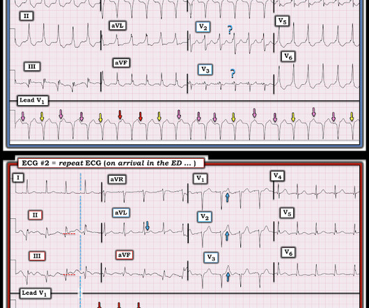
Recall that, in the setting of ACS symptoms, ST depression that are maximal in leads V1-V4 (as opposed to V5 and V6) not attributable to an abnormal QRS complex is specific for OMI. The most recent event had occurred just before being triaged. This pattern is recognizable by the ST depressions maximal in lead V4.
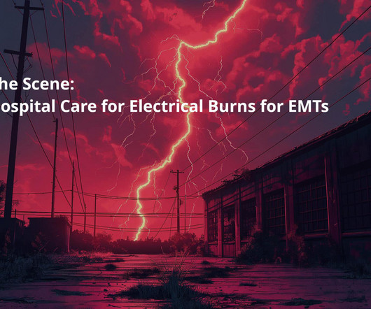
Assessing the Severity The severity of an electrical burn depends on several factors: the type of current (AC or DC), voltage, the pathway of the current through the body, the duration of contact, and the victim’s overall health. Flame Burns These are secondary burns caused when an electrical event ignites clothing or nearby materials.
The fire department, who operate at an EMT level in this municipality, arrived before us and administered 324 mg of baby aspirin to the patient due to concern for ACS. She was defibrillated and resuscitated. Takotsubo is a sudden event, not one with crescendo angina.
One must always be careful when looking for "baseline" ECGs, because the prior ECG on file may have been during another ACSevent, as this one clearly was. He was defibrillated immediately and had return of normal mental status. Cath lab activation was cancelled but the transfer was accepted for urgent cardiology evaluation.
12 minutes later, the patient went back into VFib arrest and underwent another 15 minutes of resuscitation followed by successful defibrillation and sustained ROSC. In total, he received approximately 40 minutes of CPR and 7 defibrillation attempts. That said, ACS is by far the most common and treatable cause.
I B ECG monitoring should start immediately and a defibrillator must be ready. I C Glucose-lowering therapy should be considered in ACS patients with glucose levels >10 mmol/L (>180 mg/dL), while episodes of hypoglycaemia (defined as glucose levels <_3.9 STEMI , ST-segment elevation acute myocardial infarction ).
In ACS, chest pain is the warning sign of ongoing ischemia. In this case, you should get a second defibrillator and perform double sequential external defibrillation (DSED). Simply attach a second defibrillator as shown in the diagram below and deliver max shocks from both devices simultaneously.
Several 200 J shocks did not terminate the VF, so a second defibrillator was applied for double sequential defibrillation with 400 J. She was defibrillated perhaps 25 times. After completing the ACS algorithm with amiodarone and lidocaine, there are diminishing returns on further treatments. SanzRuiz, R., Solis, J., &
He had several older ECGs on file, here are two examples: 6 days prior: 2 months prior: In the context of ACS symptoms, and when able to compare the new vs. old ECG, the top ECG is DIAGNOSTIC of OMI until proven otherwise. Defibrillation was performed, and ROSC was achieved.
After ruling out for ACS, the patient underwent angiography where he was found to have severe stable disease, which was already known. Finally Dr. Frick details how today's patient was found to have severe, stable coronary disease without evidence of an acute event. This demands an explanation -- sepsis, hemorrhage, withdrawal, etc.
We organize all of the trending information in your field so you don't have to. Join 5,000+ users and stay up to date on the latest articles your peers are reading.
You know about us, now we want to get to know you!
Let's personalize your content
Let's get even more personalized
We recognize your account from another site in our network, please click 'Send Email' below to continue with verifying your account and setting a password.


Let's personalize your content