This site uses cookies to improve your experience. To help us insure we adhere to various privacy regulations, please select your country/region of residence. If you do not select a country, we will assume you are from the United States. Select your Cookie Settings or view our Privacy Policy and Terms of Use.
Cookie Settings
Cookies and similar technologies are used on this website for proper function of the website, for tracking performance analytics and for marketing purposes. We and some of our third-party providers may use cookie data for various purposes. Please review the cookie settings below and choose your preference.
Used for the proper function of the website
Used for monitoring website traffic and interactions
Cookie Settings
Cookies and similar technologies are used on this website for proper function of the website, for tracking performance analytics and for marketing purposes. We and some of our third-party providers may use cookie data for various purposes. Please review the cookie settings below and choose your preference.
Strictly Necessary: Used for the proper function of the website
Performance/Analytics: Used for monitoring website traffic and interactions
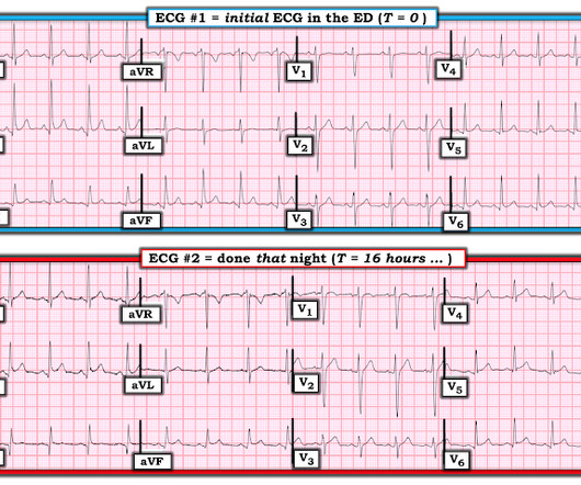
Written by Willy Frick A man in his 50s with a history of hypertension, dyslipidemia, type 2 diabetes mellitus, and prior inferior OMI status post DES to his proximal RCA 3 years prior presented to the emergency department at around 3 AM complaining of chest pain onset around 9 PM the evening prior. ECG 1 What do you think? Grines, C.
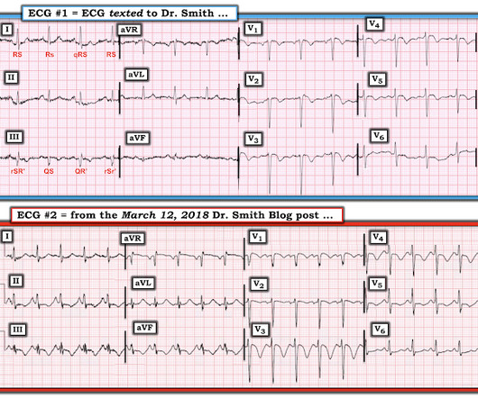
link] A 62 year old man with a history of hypertension, type 2 diabetes mellitus, and carotid artery stenosis called 911 at 9:30 in the morning with complaint of chest pain. Challenge QUESTION: The relative change in T-QRS-D is not the only thing that changes during period of time that passed between recording of the 2 ECGs shown in Figure-1.
He has already climbed Ben Nevis in Scotland, visited the Gobi desert (possibly from the comfort of his parents 4 x 4, but who’s judging) and has his bronze D of E nailed. She calls out her findings: A – OK B – 1 puncture mark to the anterior left chest wall, covered with a three-sided dressing. Actively oozing.
Angiogram No obstructive epicardial coronary artery disease Cannot exclude non-ACS causes of troponin elevation including coronary vasospasm, stress cardiomyopathy, microvascular disease, etc. IMPRESSION: 1. hours T-wave are getting larger again The patient went for an angiogram at about 7 hours after arrival. Stroke-volume:50 ml.
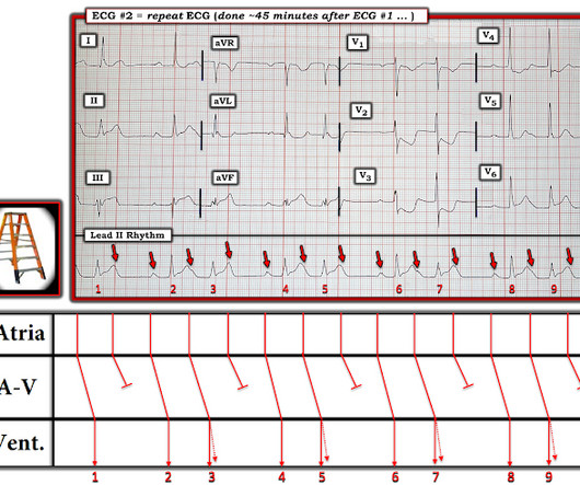
Learning Point: 1. For examples of such exceptions — See My Comment in the January 9, 2019 — August 22, 2020 — and June 30, 2023 posts in Dr. Smith's ECG Blog ). Figure-1: Comparison between the first 3 ECGs in today's case. How Would YOU Interpret the Serial Tracings in Figure-1? So they looked into the patient's chart.
A 40-something male presented with dyspnea and left arm numbness, and perhaps some chest tightness, for 11/2 hours. Therefore, this does not meet the definition of myocardial infarction ( 4th Universal Definition of MI ), which requires at least one troponin above the 99% reference range. But maybe not. Mokhtari et al.
References: 1) See this study showing an association between morphine and mortality in Non-STE-ACS: Meine TJ, Roe M, Chen A, Patel M, Washam J, Ohman E, Peacock W, Pollack C, Gibler W, Peterson E. Despite not being considered in this category, opioid medications are sometimes given for ACS. Am Heart J.
Some providers were worried about ACS because of this ECG. My answer alleviated their concern for ACS and no further workup was done for ACS. 4 important features that indicate acute right hear strain: 1. looked at consecutive patients with PE, ACS, or neither. Tachycardia (or nearly) 2. Poor R-wave progression 4.
This pattern occurs regardless of whether the cause is ACS (decreased supply) or any other cause of decreased supply or increased demand. You must understand that this pattern does not differentiate ACS from other causes of supply/demand mismatch. ST depression will not always be present in 9/12 leads — as is seen in Figure-1.
Over the last 1 week, her exertional chest pain became worse both in intensity and triggering threshold. This case fits this definition of cardiac memory. == MY Comment , by K EN G RAUER, MD ( 11/9 /2023 ): == I found Dr. What Can Sometimes Be Learned from Intermittent BBB Conduction! link] Shvilkin et al.
The pain is described as located in the midsternal area, radiating to the right arm, described as 8-9/10 and worse with deep inspirations. QRS onset is the best location for ST segment comparison, and is the location recommended by Universal Definition of MI. Figure-1: The 4 serial ECGs shown in this case ( See text ).
mm in just one lead V7-9), but as far as I can tell all of these documents specifically avoid calling this condition STEMI and specifically avoid using any terminology similar to "STEMI equivalent." I find this definition problematic because the maximal STD in posterior OMI frequently extends out to V4 rather than V3.
The fire department, who operate at an EMT level in this municipality, arrived before us and administered 324 mg of baby aspirin to the patient due to concern for ACS. mm of ST segment elevation, V2 and V3 have 1 mm of elevation, v4 has 2 mm of elevation and v5 around 1.5 Learning Points: 1. What do you think? V1 has 0.5
1 The American College of Surgeons’ (ACS) Trauma Quality Improvement Program (TQIP) Massive Transfusion in Trauma Guidelines leave a good amount of flexibility for hospitals regarding transfusion protocols, focusing more on systems-level aspects of designing and implementing MTPs.2,3 in the 1:1:1 group vs. 17.0%
The definition of massive hemoptysis is variable across publications with expectorated blood volumes ranging from 100 to 1,000 mL per 24 hours, as these volumes are difficult to estimate for any given patient. or 9 size endotracheal tube to allow for bronchoscopy and/or endobronchial blocker placement whenever necessary.
As reported within the 6th edition Manual of Emergency Airway Management, there are cardiac arrest rates between 1% and 4%, with other complications (mostly hypoxemia and hypotension) as high as 30% in patients with first-pass success [1, pg 29; 4-10]. Why is Physiologic Optimization Important? Up to 44% per other sources [12].
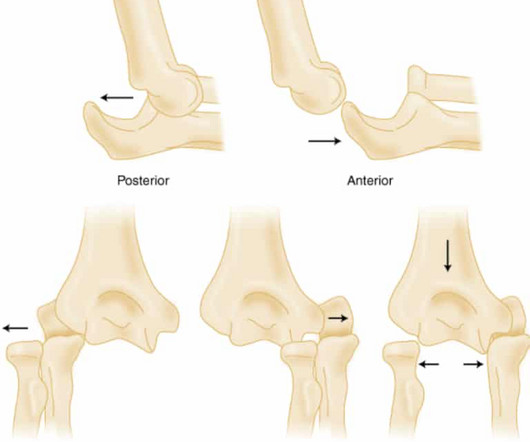
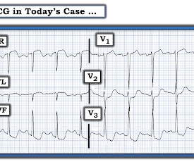
Obvious STEMI(+) OMI of inferior, posterior, and lateral walls, now with likely 2nd degree heart block type 1 (Wenckebach). STD maximal in V1-V4, without a QRS abnormality causing it, and in the setting of ACS symptoms, is posterior OMI until proven otherwise. In Figure-1 — The ST-T wave abnormality in lead V2 of ECG #1 is obvious.
study from 200 9. A large proportion of the trials come from Finland (3) and The UK (2) and also from Australia (1) and the US (1), but only a small number took place in the ED. Castro-Rodriguez JA, Beckhaus AA, Forno E. Fernandes RM, Bialy LM, Vandermeer B, Tjosvold L, Plint AC, Patel H, et al. months to 40.9
1 Indications for transplant include: Non-ischemic cardiomyopathy (49%) Ischemic cardiomyopathy (35%) Restrictive cardiomyopathy (4%) Retransplantation following failed prior transplant (3%) Hypertrophic cardiomyopathy (3%) Congenital heart disease (3%) Valvular cardiomyopathy (3%) The median survival after heart transplant is over 12 years.
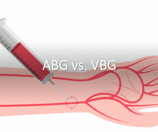
Introduction Arterial blood gas (ABG) or venous blood gas (VBG) testing is used to assess the pH and systemic carbon dioxide tension in patients, and, therefore, provide a more complete picture of their acid-base status than an isolated basic metabolic panel (BMP) (1). However, ABGs have many drawbacks compared to VBGs.
1-3 Common causes: Natural disasters such as tornadoes or earthquakes 4,5 Structure or building collapses from home fires or bombings. 6 Prolonged down time from falls, usually in the elderly Incidence is difficult to ascertain due to broad definition and that events that cause crush injuries are rather rare. 2016;20(1):135.
1 His description of cases of life-threatening infections in the perineal, genital or perianal regions were thought to be idiopathic in previously healthy men. 3-5 Fournier gangrene is a type of necrotizing soft tissue infection, which can be categorized into four types based on the infectious organism involved and other features (Table 1).
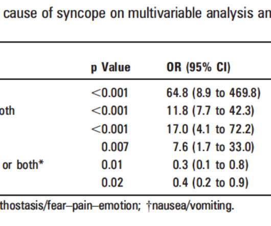
Approach to Syncope Syncope definition: Brief loss of consciousness with loss of postural tone and complete spontaneous recovery without medical intervention. Cardiac Syncope ("True Syncope") Independent Predictors of Adverse Outcomes condensed from multiple studies 1. Palpitations preceding syncope (highest value on EGSYS score) 9.
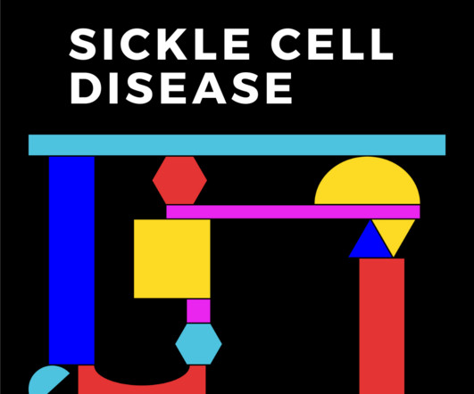
Figure 1: Clinical manifestations and long-term consequences of sickle cell disease Training and education on sickle cell disease: Training and education are crucial to improve morbidity and mortality. Investigations : Bloods show Hb of 8 g/L, White cell count 13x 10 9 /L, Platelets 570 x10 9 /L, CRP 35mg/L.
1 Life Cycle and Pathophysiology Life Cycle Humans are the only hosts for O. 1 The parasite has a 5-stage life cycle in which the blackfly acts as an obligate intermediate host. The worms can live as long as 15 years, and female worms may produce microfilariae (early-stage larvae) for up to 9 of those years. 13 Figure 1.
ED Evaluation Transport to the ED from the refugee reception center takes 1 hour. g/dL, thrombocytopenia of 96 10 9 /L, prothrombin time (PT) of 16.1 1 By the end of 2023, 117.3 million people had been forcibly displaced, representing 1 in 69 individuals or 1.5% seconds (normal 30-40 seconds), creatinine of 3.11
We organize all of the trending information in your field so you don't have to. Join 5,000+ users and stay up to date on the latest articles your peers are reading.
You know about us, now we want to get to know you!
Let's personalize your content
Let's get even more personalized
We recognize your account from another site in our network, please click 'Send Email' below to continue with verifying your account and setting a password.


Let's personalize your content