This site uses cookies to improve your experience. To help us insure we adhere to various privacy regulations, please select your country/region of residence. If you do not select a country, we will assume you are from the United States. Select your Cookie Settings or view our Privacy Policy and Terms of Use.
Cookie Settings
Cookies and similar technologies are used on this website for proper function of the website, for tracking performance analytics and for marketing purposes. We and some of our third-party providers may use cookie data for various purposes. Please review the cookie settings below and choose your preference.
Used for the proper function of the website
Used for monitoring website traffic and interactions
Cookie Settings
Cookies and similar technologies are used on this website for proper function of the website, for tracking performance analytics and for marketing purposes. We and some of our third-party providers may use cookie data for various purposes. Please review the cookie settings below and choose your preference.
Strictly Necessary: Used for the proper function of the website
Performance/Analytics: Used for monitoring website traffic and interactions
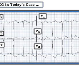
Many of the changes seen are reminiscent of LVH with “strain,” and downstream Echo may very well corroborate such a suspicion, but since the ECG isn’t the best tool for definitively establishing the presence of LVH, we must favor a subendocardial ischemia pattern, instead. He awoke earlier that morning in his usual state of health.
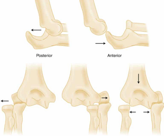
Elbow Dislocation Definition: Disarticulation of the proximal radius & ulna bones from the humerus Epidemiology: Incidence Second most common joint dislocation (after shoulder) in adults Most commonly dislocated joint in children Accounts for 10-25% of all injuries to the elbow ( Cohen 1998 ) Posterolateral is the most common type of dislocation (..)
In an attempt to clarify language, a consensus definition was developed. The definition requires the following three components: An end QRS notch (sometimes called a J wave) or slur, in the case of a slur it must lie entirely above the isoelectric baseline The peak amplitude of the notch or slur should be ≥ 0.1 Back to the case.
Then assume there is ACS. Confirmation of sinus tachycardia should be easy to verify when the heart rate slows a little bit ( as the patient's condition improves ) — allowing clearer definition between the T and P waves. The ST depression usually resolves, or is clearly resolving (getting much better).
These results are not definitive, but considering the rarity of demyelination, and the magnitude of the mortality results, this should probably influence clinical practice until we get the proper RCTs. Welcome to the first episode of the Broomedocs podcast for 2025. Listen in and learn! WOMAN are so negative WOMAN-2 Trial Collaborators.
RBBB + LAFB in the setting of ACS is very bad. Some patients have baseline RBBB with LAFB, but in patients with likely ACS, these are associated with severe infarction with cardiac arrest, cardiogenic shock or impending shock. Patients with ACS and RBBB/LAFB usually have a left main vs. proximal LAD. There is STE in aVR.
REBEL Cast Ep114 – High Flow O2, Suspected ACS, and Mortality? PMID: 33653685 Clinical Question: Is there an association between high flow supplementary oxygen and 30-day mortality in patients presenting with a suspected acute coronary syndrome (ACS)? Click here for Direct Download of the Podcast Paper: Stewart, RAH et al.
The ECG does not show any definite signs of ischemia. He denied any exertional chest pain. The below ECG was recorded. It is unclear if the patient was pain free at this time. In fact, the ECG was described as normal, and without serial ECGs or prior ECGs for comparison it could be.
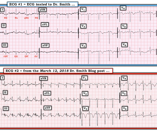
This was texted to me from a former resident, while working at a small rural hospital, with the statement: "I can’t convince myself of anything here, but he’s a 63-year-old guy with prior stents and a good story for ACS." Chest pain or discomfort) What do you think? Here was my response: "Suspicious for inferior posterior OMI.
Category 1 : Sudden narrowing of a coronary artery due to ACS (plaque rupture with thrombosis and/or downstream showering of platelet-fibrin aggregates. Smith : This is ACS even if the troponin returns normal, and the first troponin especially might return normal. This results in Type I MI. This results in Type II MI. Severe HTN d.
There were zero patients in this study with a "normal" ECG who had any kind of ACS! This defies all previous data on acute MI which would show that even undetectable troponins do not have a 100% negative predictive value. So this study is actually worthless. Deutch et al. West J Emerg Med 2024).
1, 2] The most clinically useful definition to account for this entire constellation is intraventricular conduction delay. Anecdotally, had there been symptoms unequivocally consistent with ACS then one could justifiably make the case for a potential D1 occlusion. He denied difficulty breathing, epigastric pain, or chest discomfort.
Click here for Direct Download of the Podcast Paper: Aykan AC et al. Because the lungs receive 100% of cardiac output, it has been hypothesized that a lower dose of thrombolytic therapy may still be effective with a better safety profile [3][4]. REBEL Cast Ep123: Reduced-Dose Systemic Peripheral Alteplase in Massive PE?
This is diagnostic of ACS; it appears to be a reperfused acute inferior OMI. Suppose the OMI had been recognized, or suppose another ECG had been recorded and it showed definite OMI. He reports feeling nauseated with emesis. He denies taking aspirin or antihypertensive medications for the last year and a half.
Angiogram No obstructive epicardial coronary artery disease Cannot exclude non-ACS causes of troponin elevation including coronary vasospasm, stress cardiomyopathy, microvascular disease, etc. It is not yet available, but this is your way to get on the list. link] Case continued She arrived in the ED and here is the first ED ECG.
Thus, this does NOT meet STEMI criteria (though, as of 2022, it is a formal "STEMI equivalent", assuming everyone agrees that this is de Winter morphology, for which there is currently no objective definition). Everywhere I'm aware of (only about 5 systems), the ECGs are not even recorded in the "STEMI database." What a farce.
Therefore, this does not meet the definition of myocardial infarction ( 4th Universal Definition of MI ), which requires at least one troponin above the 99% reference range. You can see the deficiency of the definition of MI. Because there is reciprocal ST depression in aVL, this should not be called early repol. But maybe not.
Because the most severe LAD OMIs can cause ischemic failure of the RBB and LAF, any patient with ACS symptoms and new RBBB and LAFB with any concordant STE has LAD OMI until proven otherwise. Post cath EF was estimated at 15% with severe global hypokinesis, and akinesis of the apex. Long term outcome is unavailable.
80%) and definitely too much for hour to hour. However, the Definition of MI requires at least one value above the 99th percentile, which for a male is 34 ng/L (16 ng/L for women). Thus, these troponins are very concerning for ACS, and subsequent ones will probably be diagnostic of acute MI. His angiogram is shown below.
Because there was proven thrombus (ACS) but the troponin never went above the 99% reference range (and therefore cannot be called MI -- definition of MI requires rise and/or fall of troponin with at least one value above the 99% reference range), this is UNSTABLE ANGINA with ST Elevation. Fortunately, that is exactly what happened.
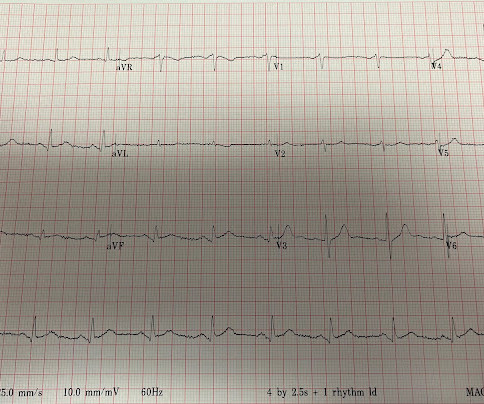
Similarly, the OMI paradigm respects ACS as a dynamic process in which ECG changes reflect the phase of myocardial injury and risk stratify which patients may benefit from emergent PCI. Although tiny in size — the BLUE arrows highlight definite ST elevation in leads I and aVL. In fact, use of antidyrhythimcs (e.g., Leave it alone.
However , this patient is having chest discomfort, and by definition then she should be considered not to be stable. Ischemia from ACS causing the chest discomfort, with VT another consequence (or coincidence)? Is this: 1. In either case, prompt cardioversion is indicated. In either case, prompt cardioversion is indicated.
Episode 107: Eclampsia Definition: Severe hypertensive disease of pregnancy (HDP) with new onset tonic-clonic, focal, or multifocal seizures or unexplained altered mental status in a patient who is pregnant or postpartum and there’s no other causative etiologies. Today on the emDOCs cast with Brit Long ( @long_brit) , we cover eclampsia.
There is definite reperfusion. There was definite evolution of the ECG. He had been smoking an opiate and suddenly collapsed. He was ventilated with BVM on arrival. He awoke with naloxone. This EKG was recorded as part of a standing order for critical care. He denied any CP or SOB. Maybe there is some spontaneous reperfusion?
References: 1) See this study showing an association between morphine and mortality in Non-STE-ACS: Meine TJ, Roe M, Chen A, Patel M, Washam J, Ohman E, Peacock W, Pollack C, Gibler W, Peterson E. Link to abstract Link to full text 2) Use of Morphine in Non-STE-ACS is independently associated with mortality, at odds ratio of 1.4
ACS then becomes less likely. BOTTOM Line: There clearly is enough on this initial ECG to support Dr. Nossen's concern that a definitive diagnosis needed to be made on this young man with new, persistent chest pain. On arrival patient was slightly tachycardic. HR about 90-100/min. Other vital signs normal. How did the Queen do?
When Pendell and I are coding ECGs for the Queen's training, this is one category: "Definite ischemia, difficult to differentiate between posterior OMI and subendocardial ischemia." In our opinion it should not be given in ACS unless you are committed to the cath lab. He was diagnosed as NSTEMI.
But because the patient had no chest pain or shortness of breath, it was not deemed to be from ACS. But because the patient had no chest pain or shortness of breath, it was not deemed to be from ACS. They were less likely to have STEMI on ECG, and more likely to be initially diagnosed as non-ACS. Potassium was normal.
Despite the seemingly worrisome ST-T wave changes on serial tracings shown in Figure-1 — an acute event was definitively ruled out by 4 consecutive negative hs troponins — with further support provided by an Echo showing excellent LV function without wall motion abnormality. So they looked into the patient's chart. Learning Point: 1.
This week we’re looking at the other ACS, the surgical ACS, the old abdominal compartment syndrome. This week we’re looking at the other ACS, the surgical ACS, the old abdominal compartment syndrome. This week we’re looking at the other ACS, the surgical ACS, the old abdominal compartment syndrome.
ACS surgeons appeared to select surgery as their initial choice substantially more frequently than other subspecialties. ACS surgeons would have sent 6/43 patients for ERCP or MRCP (14%), whereas surgical oncologists would have sent a higher percentage of patients for ERCP or MRCP (7/18 or 38.9%). and specificity of 88.0%
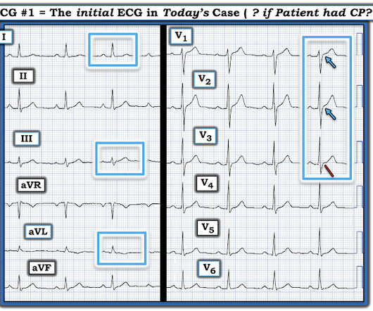
Definition: Technically, for the tracing in ECG #1 to truly “qualify” as Wellens’ Syndrome — there should be no CP at the time the ECG is recorded. However, in the chest leads — there has definitely been evolution , in that ST segments are more coved and slightly more elevated in virtually all chest leads. No diaphoresis or vomiting.
He had no symptoms of ACS. His HEAR score (before troponin resulted) was documented at 3, with documentation stating "low suspicion for ACS." A troponin this high in a patient with no known chronic troponin elevation, and active acute ACS symptoms, has a very high likelihood of type 1 ACS regardless of the ECG.
You ask your anaesthetist to get ready to sedate or intubate depending on their status – Significant risk to the department – you make sure security is aware And your patient arrives. Ranulf is quite a sweet, round-faced boy, accompanied by his traumatised-looking mother as he is wheeled to your trauma bay.
There is perhaps a tiny J-wave in several of the QRS complexes in V3, but it would not be enough to definitively say there is a J-wave. Lead aVL, for example, has a definite J-wave. More importantly, this patient has either no chest pain at the time of this ECG or greatly diminished chest pain. Stat echo would also be helpful.
If you like the types of questions we answer on The Curious Clinicians, you’ll definitely want to check that out and subscribe. Listen to the episode [link] Credits & Citatio n Episode written by Avi Cooper Show notes written by Giancarlo Buonomo and Avi Cooper Audio edited by Clair Morgan of nodderly.com Cooper AZ, Breu AC, Abrams HR.
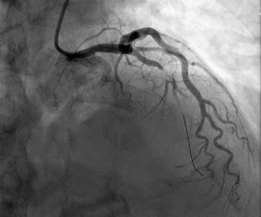
Here is an article I wrote: Updates on the ECG in ACS. Was this: 1) ACS with ischemia and spontaneous reperfusion? The patient was taken for an angiogram and had an 80% LAD lesion, but it could not be definitely determined whether this was an acute thrombotic lesion or a chronic stable lesion. See image with lines below).
Apple Podcasts , Spotify , Listen Here Resuscitative endovascular balloon occlusion of the aorta (REBOA) is a minimally invasive way of providing resuscitative aortic occlusion in severe hemorrhage to gain temporary hemorrhage control as a bridge to definitive procedures. ” As a result, Jansen et al. ” As a result, Jansen et al.
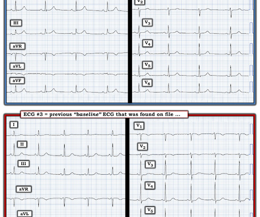
3) RV Failure leads to hypotension but NOT pulmonary edema (unlike LV failure) 4) Repeat ECGs, right sided ECG and bedside echo may be helpful in making a diagnosis of ACS. Lead aVL is definitely not normal. Any cause of pulmonary hypertension. At this point an old ECG on file was found for comparison.
The patient has ACS by history, active pain, and an elevated troponin. By definition , this is acute myocardial infarction, the only question now is the etiology. It is impossible to overstate the importance of putting the ECG and troponin into the context of the clinical history. Smith : at this point, the ECG becomes irrelevant.
Some providers were worried about ACS because of this ECG. My answer alleviated their concern for ACS and no further workup was done for ACS. showed that , when T-waves are inverted in precordial leads, if they are also inverted in lead III and V1, then pulmonary embolism is far more likely than ACS. What is an S1Q3T3?
See this study showing an association between morphine and mortality in ACS: Use of Morphine in ACS is independently associated with mortality, at odds ratio of 1.4. Do NOT give it unless you are committed to the cath lab!! Cath attending is aware. I think it is OMI; am I right? See this case: A man his 50s with chest pain.
This pattern occurs regardless of whether the cause is ACS (decreased supply) or any other cause of decreased supply or increased demand. You must understand that this pattern does not differentiate ACS from other causes of supply/demand mismatch. A "STEMI alert" was called and soon cancelled.
Moreover, what I call "domed" T-wave inversion in V1-V3 is typical for acute PE and NOT typical of ACS (i.e., Moreover, T-wave inversion in V1-V3 due to ACS is typically seen in reperfusion states when the patient is symptom free. Wellens'), presumably because the source of the T-wave inversion is RV strain, not LV ischemia.
We organize all of the trending information in your field so you don't have to. Join 5,000+ users and stay up to date on the latest articles your peers are reading.
You know about us, now we want to get to know you!
Let's personalize your content
Let's get even more personalized
We recognize your account from another site in our network, please click 'Send Email' below to continue with verifying your account and setting a password.


Let's personalize your content