This site uses cookies to improve your experience. To help us insure we adhere to various privacy regulations, please select your country/region of residence. If you do not select a country, we will assume you are from the United States. Select your Cookie Settings or view our Privacy Policy and Terms of Use.
Cookie Settings
Cookies and similar technologies are used on this website for proper function of the website, for tracking performance analytics and for marketing purposes. We and some of our third-party providers may use cookie data for various purposes. Please review the cookie settings below and choose your preference.
Used for the proper function of the website
Used for monitoring website traffic and interactions
Cookie Settings
Cookies and similar technologies are used on this website for proper function of the website, for tracking performance analytics and for marketing purposes. We and some of our third-party providers may use cookie data for various purposes. Please review the cookie settings below and choose your preference.
Strictly Necessary: Used for the proper function of the website
Performance/Analytics: Used for monitoring website traffic and interactions
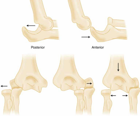
Elbow Dislocation Definition: Disarticulation of the proximal radius & ulna bones from the humerus Epidemiology: Incidence Second most common joint dislocation (after shoulder) in adults Most commonly dislocated joint in children Accounts for 10-25% of all injuries to the elbow ( Cohen 1998 ) Posterolateral is the most common type of dislocation (..)
We documented that the majority of stenotic lesions had compensatory enlargement and thus exhibited remodeling. Strictly speaking — this case does not at this time qualify as MINOCA — because the negative cath and 2 normal hs-troponins done 6 hours apart failed to document infarction. (
Cardiology consult note written around that time documents that "Pain improved with NTG, morphine in ED but still present." As a result, even before looking at this patient's initial ECG — he falls into a high -prevalence likelihood group for ACS ( for an A cute C oronary S yndrome ). Repeat cTnI drawn at around 8 AM was 3.910 ng/mL.
REBEL Cast Ep114 – High Flow O2, Suspected ACS, and Mortality? PMID: 33653685 Clinical Question: Is there an association between high flow supplementary oxygen and 30-day mortality in patients presenting with a suspected acute coronary syndrome (ACS)? Click here for Direct Download of the Podcast Paper: Stewart, RAH et al.
showed that , when T-waves are inverted in precordial leads, if they are also inverted in lead III and V1, then pulmonary embolism is far more likely than ACS. In this study, (quote) "negative T waves in leads III and V 1 were observed in only 1% of patients with ACS compared with 88% of patients with Acute PE (p less than 0.001).
Sponsor Freed is an AI scribe that listens, transcribes, and writes medical documentation for you. It turns clinicians’ patient conversations into accurate documentation – instantly. Some of these cascades, even if cumbersome to the patient, make a certain amount of physiological sense. However, one cascade is a little less obvious.
Triage documented a complaint of left shoulder pain. Smith : As Willy states, ACS with persistent symptoms is a guideline recommended indication for <2 hour angio (both ACC/AHA and ESC). Smith : As Willy states, ACS with persistent symptoms is a guideline recommended indication for <2 hour angio (both ACC/AHA and ESC).
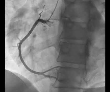
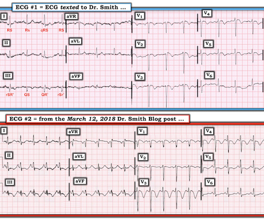
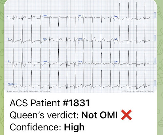
The documentation does not describe any additional details of the history. They also documented "Reproducible chest tenderness." Thus, these troponins are very concerning for ACS, and subsequent ones will probably be diagnostic of acute MI. The following ECG was obtained. ECG 1 What do you think? His angiogram is shown below.
Notoriously elusive, with a high misdiagnosis rate, thoracic aortic dissection (AD) can mimic many conditions, including acute coronary syndrome (ACS, the most common), gastroesophageal reflux disease (GERD), stroke, and spinal-cord compression. The patient is admitted for ACS to a cardiologist who says he will see the patient in the morning.
Click here for Direct Download of the Podcast Paper: Aykan AC et al. Because the lungs receive 100% of cardiac output, it has been hypothesized that a lower dose of thrombolytic therapy may still be effective with a better safety profile [3][4]. REBEL Cast Ep123: Reduced-Dose Systemic Peripheral Alteplase in Massive PE?
Article: Vaeli Zadeh A, Wong A, Crawford AC, Collado E, Larned JM. References: Vaeli Zadeh A, Wong A, Crawford AC, Collado E, Larned JM. Guideline-based and restricted fluid resuscitation strategy in sepsis patients with heart failure: A systematic review and meta-analysis [published online ahead of print, 2023 Aug 9]. 2.89, p = 0.01.
The patient was thought to have low likelihood of ACS, and cardiology recommended repeat troponin, urine drug testing, and echocardiogram. At that point, cardiology elected to treat for ACS. The operator documented thoughtful consideration of risks and benefits of stent placement. Initial hscTnI was 10 ng/L (ref. <14).
Click here to sign up for Queen of Hearts Access Given the lack of intracranial hemorrhage, the patient was administered aspirin for suspected ACS and cardiology was consulted. Preliminary findings documented in the cath lab were “Anterior STEMI and no significant coronary artery disease.” (!!!) or basilar ischemia. ng/mL and 0.10
The Eastern Association for the Surgery of Trauma (EAST) , the National Association of EMS Physicians (NAEMSP) , and the American College of Surgeons Committee on Trauma (ACS-COT) all support the recommendation against the use of spinal immobilization in patients with isolated penetrating injuries. to prevent movement of the spine.
None of the patients were documented to have joint disease at follow up. When you read the Morsel on Perichonditis of the ear last week ( or perhaps the Plantar Puncture Morsel from many many many weeks ago ) you may have objected because of the mention that, when indicated, fluroquinolones are safe in children.
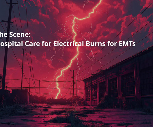
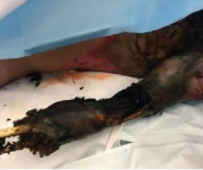
Assessing the Severity The severity of an electrical burn depends on several factors: the type of current (AC or DC), voltage, the pathway of the current through the body, the duration of contact, and the victim’s overall health. The entry and exit wounds are key indicators, but they can be small or hidden under clothing.
While one may argue that nitro really has no mortality benefit in ACS, I have seen patients with CHF present with hyper tension and inferior S-T elevation, in which the providers were scared to even look at the bottle of nitro. References Kimbrell J, Kreinbrook J, Poke D, Kalosza B, Geldner J, Shekhar AC, Miele A, Bouthillet T, Vega J.
He had no symptoms of ACS. His HEAR score (before troponin resulted) was documented at 3, with documentation stating "low suspicion for ACS." A troponin this high in a patient with no known chronic troponin elevation, and active acute ACS symptoms, has a very high likelihood of type 1 ACS regardless of the ECG.
Cardiology documents their interpretation of ECG in their consult note - “atrial paced with old LBBB” The patient stayed at outside hospital (which does not have cardiac cath capabilities). The cath report showed: Significant stenosis with subtotal occlusion (99%) in the prox to mid Lcx, culprit of ACS, TIMI flow 1.
Ongoing pain noted throughout all documentation, but after nitro drip and prn morphine, "pain improved to 2/10." References: 1) See this study showing an association between morphine and mortality in Non-STE-ACS: Meine TJ, Roe M, Chen A, Patel M, Washam J, Ohman E, Peacock W, Pollack C, Gibler W, Peterson E. Repeat trop 150 ng/L.
Because the most severe LAD OMIs can cause ischemic failure of the RBB and LAF, any patient with ACS symptoms and new RBBB and LAFB with any concordant STE has LAD OMI until proven otherwise. In EMS2 ECG, the T waves in V5 is possibly hyperacute. Post cath EF was estimated at 15% with severe global hypokinesis, and akinesis of the apex.
ACS surgeons appeared to select surgery as their initial choice substantially more frequently than other subspecialties. ACS surgeons would have sent 6/43 patients for ERCP or MRCP (14%), whereas surgical oncologists would have sent a higher percentage of patients for ERCP or MRCP (7/18 or 38.9%). and specificity of 88.0%
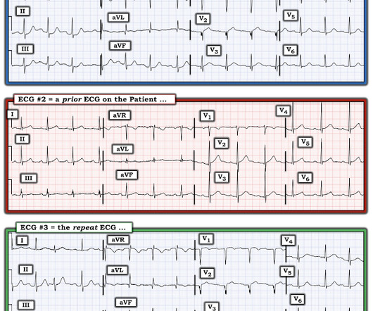
He had no previously documented medical problems except polysubstance use. Similarly, the OMI paradigm respects ACS as a dynamic process in which ECG changes reflect the phase of myocardial injury and risk stratify which patients may benefit from emergent PCI. An ECG was obtained shortly after arrival: What do you think?
Type 1 is the acute deterioration in kidney function seen in cardiogenic shock from ACS. This segues relatively nicely into a section of the document on palliative care. Type 1 is the acute deterioration in kidney function seen in cardiogenic shock from ACS. To start with there are apparently 5 types of cardiorenal syndrome.
After rethinking the case, he remained concerned about ACS and subsequently performed a point-of-care ultrasound in order to evaluate for regional wall motion abnormality. We assume that at some point the patient's pain returned, but it is not documented, so exactly when this happened is uncertain. There is no age cut-off for ACS.
Ischemia from ACS causing the chest discomfort, with VT another consequence (or coincidence)? Cardioversion will address the rhythm problem immediately, also if the chest discomfort subsides when SR is restored, ischemia from ACS becomes much less likely. In either case, prompt cardioversion is indicated.
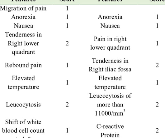
It is worth doing and documenting serial abdomen exams in non-specific abdo pains. References Meltzer AC, Baumann BM, Chen EH, Shofer FS, Mills AM. Abdominal pain is a common ED presentation and one of the top differential for RLQ pain is Acute Appendicitis. The original Alvarado score was on a 10 point scale.
The AHA/ACC guidelines recommend emergent cardiac catheterization for patients with concern for ACS and refractory chest pain despite maximum medical therapy defined as aspirin + clopidogrel/ticagrelor + heparin/enoxaparin. link] He was admitted to the cardiology unit for serial troponin measurements and concern for possible ACS.
Some providers were worried about ACS because of this ECG. My answer alleviated their concern for ACS and no further workup was done for ACS. showed that , when T-waves are inverted in precordial leads, if they are also inverted in lead III and V1, then pulmonary embolism is far more likely than ACS. What is an S1Q3T3?
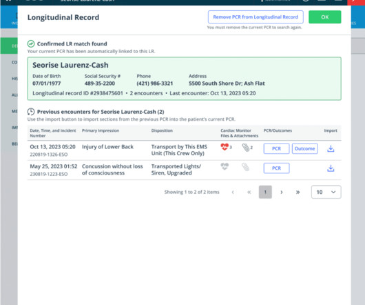
This new feature breaks down the data silos that are inherent in typical prehospital documentation and changes the paradigm of patient care documentation from encounter-based to patient-centric. You’ll now have a holistic view of frequent patients, along with the ability to view previous 12-leads, PCRs and HDE Outcomes.
Current can be alternating current (AC) or direct current (DC) with AC typically more dangerous as it is more likely to cause tetanic contractions and increase contact time with the electrical source. 2,3,5 Except for laundry or electrical car outlets (240 V AC), all U.S. household outlets are rated at 120 V AC.
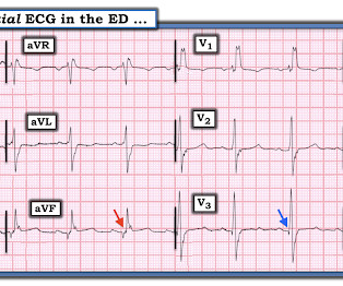
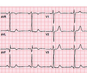
But there are also hyperacute T waves (HATW) in V4-5, which exclude early repolarization and pericarditis, leaving only LAD occlusion for this patient presenting with classic symptoms of ACS. What do you think? What do you think? Theres normal sinus rhythm with normal conduction, right axis and delayed R wave, and normal voltages.
It is easy to say this in retrospect, especially not being the one in charge of this overcrowded waiting room full of unseen patients, but an elderly patient with known CAD and ongoing ACS-sounding chest pain despite medical management with positive troponin is already an indication for emergent cath, regardless of the ECG!
If it is maximal in V1-V4, and the patient's presentation in consistent with ACS (as this certainly is), then it is DIAGNOSTIC of Occlusion with 90% specificity (We have an upcoming article that proves this). Angiogram: "ACS - Non ST Elevation Myocardial Infarction. This was not recognized. The patient was started on a nitro drip.
There is a substantial ANZICS document on tracheostomy that forms the structure for this tasty morsel. doi: 10.21037/acs.2018.03.01. Intensivists have embraced the tracheostomy as an ICU procedure. It’s one of the most invasive and one of the riskier procedures we do. Surgical anatomy of the trachea. Ann Cardiothorac Surg.
Since ACS is so dynamic, with thrombi forming and lysing continuously, and because the ECG and angiogram are rarely simultaneous, it is probable that de Winter's T-waves are recorded in a window when the artery is barely open. This is the longest lasting I have ever documented a hyperacute T wave without going "up" or "down."
But it does prove that the patient has coronary disease and makes the probability that his chest pain is due to ACS very very high. It is proven better than angiography alone in stable angina , and also has been shown to improve decisions on stenting non-culprit lesions in ACS. We need to do some more investigation.
I did not think it was due to ACS, but we ordered an ED ECG immediately: What do you think? But in this case the clinical scenario is not right for acute ACS with OMI, and there is very high voltage, and the patient is very young, (though beware of young patients , even 29 year olds!! There is profound "inferior" ST Depression.
There have been documented cases of overdose, and of note, there is no known antidote. Given the fact that he has not had these headaches before and has diffuse symptoms including weakness, lab work and head imaging are obtained. There were no acute findings on head CT.
As he documented, “This patient is experiencing chest pain consistent with an acute coronary syndrome. As cardiology documented, “possible STEMI. Most importantly , while waiting for the paradigm to evolve, maintain focus on our true goal for our patients with ACS: to identify and reperfuse patients with acute occlusion MI.”
We can see that there is evolution of the elevations, worsening reciprocal change, as well as evolution of posterior involvement (right precordial R-waves with ST depression) There was very little documentation surrounding these ECGs. Beware of ACS presenting with atypical symptoms, including absence of chest pain.
It is true that other documents occasionally describe "abnormal ST segment elevation" in the posterior leads (commonly accepted criteria is 0.5 mm in just one lead V7-9), but as far as I can tell all of these documents specifically avoid calling this condition STEMI and specifically avoid using any terminology similar to "STEMI equivalent."
If a patient presents with symptoms of ACS, has an elevated troponin, and has persistent symptoms in spite of medical therapy [antiplatelet, antithrombotic, and anti-ischemic (nitro)], then cath lab activation is indicated regardless of ECG findings. 99% of inferior OMI are either obvious or have some amount of ST depression in aVL.
We organize all of the trending information in your field so you don't have to. Join 5,000+ users and stay up to date on the latest articles your peers are reading.
You know about us, now we want to get to know you!
Let's personalize your content
Let's get even more personalized
We recognize your account from another site in our network, please click 'Send Email' below to continue with verifying your account and setting a password.


Let's personalize your content