This site uses cookies to improve your experience. To help us insure we adhere to various privacy regulations, please select your country/region of residence. If you do not select a country, we will assume you are from the United States. Select your Cookie Settings or view our Privacy Policy and Terms of Use.
Cookie Settings
Cookies and similar technologies are used on this website for proper function of the website, for tracking performance analytics and for marketing purposes. We and some of our third-party providers may use cookie data for various purposes. Please review the cookie settings below and choose your preference.
Used for the proper function of the website
Used for monitoring website traffic and interactions
Cookie Settings
Cookies and similar technologies are used on this website for proper function of the website, for tracking performance analytics and for marketing purposes. We and some of our third-party providers may use cookie data for various purposes. Please review the cookie settings below and choose your preference.
Strictly Necessary: Used for the proper function of the website
Performance/Analytics: Used for monitoring website traffic and interactions
Our experience: Traditionally, ED physicians do not like ordering urine drug screens (UDS). We have certainly seen patients who have pain which is controlled and still have psychomotor agitation and sympathetic activation, leading some to require ICU admission for dexmedetomidine and/or ketamine infusion. 2023 Aug 1;89(2):231.
European Journal of Internal Medicine , [link] You can listen to my 27-minute rant on Youtube here: [link] This multinational trial looked at a three-pronged diagnostic protocol in the ED for adults with suspected acute aortic syndromes. The protocol used the ADD score, a POCUS echo protocol and D-dimer to try and exclude AAS in the ED.
The patient was upgraded to the ICU for closer monitoring. showed that , when T-waves are inverted in precordial leads, if they are also inverted in lead III and V1, then pulmonary embolism is far more likely than ACS. looked at consecutive patients with PE, ACS, or neither. Kosuge et al. Witting et al. of controls.
However, RSI has never been shown to reduce the risk of aspiration in the ED (13) or during emergent OR cases (14). While RSI should remain the gold standard in the vast majority of patients in the ED, FI presents an additional technique to mitigate anatomic or physiologic risk. To date, ketamine has been the agent of choice (12).
Ischemia from ACS causing the chest discomfort, with VT another consequence (or coincidence)? Cardioversion will address the rhythm problem immediately, also if the chest discomfort subsides when SR is restored, ischemia from ACS becomes much less likely. This patient presented to the ED “after a couple of days of chest discomfort”.
Moreover, the Queen is only supposed to be used with a high pretest probability of ACS/OMI. The patient was admitted to the ICU for close monitoring and electrolyte repletion and had an uneventful hospital course. We just finished training version 2 with some cases of hypokalemia, so that is in the future. As per Drs.
It has been well over a year since the controversial publication of the Agency for Healthcare Research and Quality (AHRQ) report on diagnostic errors in the emergency department (ED). percent of ED visits resulted in preventable death as result of diagnostic error. Further diagnostic testing in the ICU identified salicylate toxicity.
The patient vomited once and given the more intense pain decided to come to the ED. Smith comment: We have shown that use of opiates is associated with worse outcomes in ACS: Bracey, A. 2-hour hsTn: 615 ng/L; bedside ED echo (without contrast) did not show a clear wall motion abnormality (WMA). Abstract 556.
All you know, back in ED, is that the ETA is 10 minutes, and there is a single stab wound to the chest. The ODP is caught up leaving theatres and has not yet made it down to ED. They found NO difference in drain failure rates ( 11% pigtail vs 13% chest tube P=0.74), total daily volume drained or length of ICU stay between groups.
The NIHSS cutoff that predicts outcomes is 4 points higher in AC compared with PC infarctions. Median time from ED arrival to diagnosis was 8 hours 24 min in one study, with only 19% being diagnosed within the 4.5-hour Post TW, ed. NIHSS does have limitations when applied to posterior circulation (PC) strokes. Neurohospitalist.
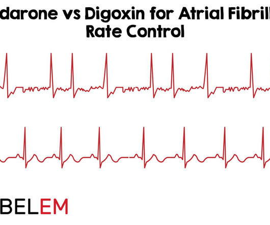
Background Information: Atrial fibrillation with rapid ventricular rate (RVR) is one of the many tachydysrhythmias we encounter in the Emergency Department (ED). 2 Amiodarone is commonly known for its anti-arrhythmic properties and a commonly used agent in the Intensive Care Unit (ICU).
1 The American College of Surgeons’ (ACS) Trauma Quality Improvement Program (TQIP) Massive Transfusion in Trauma Guidelines leave a good amount of flexibility for hospitals regarding transfusion protocols, focusing more on systems-level aspects of designing and implementing MTPs.2,3 ACS TQIP Best Practice Guidelines. 248(3):447-58.
She did not receive any opioids (which would mask her pain without affecting any underlying ACS). She was asymptomatic at the time of this ECG recorded on arrival to our ED: What do you think? By the time the patient arrived at our facility, she had received aspirin and nitroglycerin, and her pain had apparently completely resolved.
Written by Pendell Meyers A man in his late 40s with several ACS risk factors presented with a chief complaint of chest pain. The cardiologists felt that the ECG did not represent ACS, and thought it was more likely pericarditis, so they did not take him to the cath lab. in the ICU but survived with excellent function.
This single-centre academic urban institution in the United States (US) undertook a 10-year retrospective observational study of paediatric intubation and bougie use in their emergency department (ED). During a 6-month period, paediatric patients (< 18 years old) who underwent tracheal intubation in their ED were included in the study.
One of the most hair-raising presentations to the emergency department (ED) can be massive hemoptysis with respiratory failure. References Deshwal H, Sinha A, Mehta AC. 6 Position the patient with the head of the bed at 30 to 45 degrees during intubation whenever possible, and use an 8.5 A special thanks to Drs. 2021;42(1):145-159.
An example using a real case I had while on call in the ICU: A 61-year-old female had a post-induction arrest on the wards/hospital telemetry floor after being intubated for airway protection. PMID: 30060961 Koller AC, et al. In a PCAC 1 or 2, we may prioritize a cath and tolerate a couple hours without ICU Neuroresuscitation.
This is her pre-hospital ECG: This is her first ECG in the ED: What do you think? She also received an additional nitro in the ED after receiving aspirin and nitro via EMS. I took part in her ICU care and she was extubated and stable to transfer to a stepdown unit after a few days. Case A 30 something y.o.
The patient was managed in the ICU and had serial troponins. An angiogram confirmed ACS as the etiology. Figure-1: The first 2 ECGs shown in this case ( See text ). == C OMMENT : As per Dr. Smith — E CG # 1 was the initial tracing on this patient who presented to the ED already intubated for respiratory failure. First was 2.9
A 64-year-old male presents by EMS to the ED with shortness of breath. 1 There are over 50,000 visits related to heart transplant in the United States each year and over half of these patients are admitted to the hospital from the ED. We’ll keep it short, while you keep that EM brain sharp.
As the only respiratory therapist in the ED has been paged and is starting BiPAP for this patient, an overhead call for two incoming trauma alerts from a multivehicle collision sounds. Because the RT responsible for drawing arterial blood gases is busy caring for these patients, ABGs will be delayed.
An 8-year old male with a history of sickle cell anemia presents to the ED for evaluation of fever for 2 days and “feeling like I can’t get a full breath”. 768: Epidemiology of Hospital Based ED Visits due to Sickle Cell Crisis and Acute Chest Syndrome in Kids. C or 100.4 mg/kg, max 4 mg per dose q20-30min) or hydromorphone (0.01-0.02
F, HR 48, RR 28, BP 104/62, SPO2 88% on non-rebreather mask The patient’s friend who brought her to the ED tells you the patient made suicidal statements earlier in the day and was found in her yard shed. These are send-out labs with turn-around times that make them unlikely to affect the ED course or guide treatment. Toxicology.
Haematology specialist clinics are key to manage the chronic side of the disease, while ED doctors should be able to act rapidly on the common acute emergencies. with thanks A 15-month-old Kenyan boy presents to ED with right hand swelling. A 10-year-old boy with known SCA presents to ED due to severe pain in the legs.
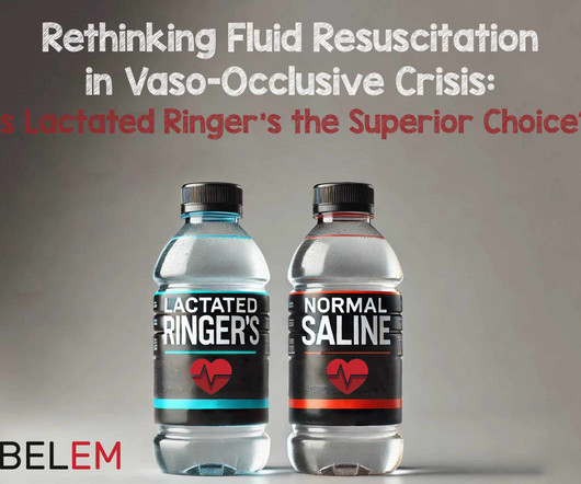
Paper: Alwang AK, Law AC, Klings ES, Cohen RT, Bosch NA. The characteristics that were significantly different between the LR and NS exposure groups race, organ dysfunction at presentation, ICU admission, hemoglobin SS genotype, discharge year, and hydroxyurea use were appropriately included as confounders in the TMLE analysis.
ED Evaluation Transport to the ED from the refugee reception center takes 1 hour. Labs Laboratory workup in the ED is notable for a leukocytosis of 41,000/L, hemoglobin of 6.5 She is sent to the medical ward after three days in the ED with the diagnoses of resolving septic shock, severe malaria, and AKI.
We organize all of the trending information in your field so you don't have to. Join 5,000+ users and stay up to date on the latest articles your peers are reading.
You know about us, now we want to get to know you!
Let's personalize your content
Let's get even more personalized
We recognize your account from another site in our network, please click 'Send Email' below to continue with verifying your account and setting a password.


Let's personalize your content