This site uses cookies to improve your experience. To help us insure we adhere to various privacy regulations, please select your country/region of residence. If you do not select a country, we will assume you are from the United States. Select your Cookie Settings or view our Privacy Policy and Terms of Use.
Cookie Settings
Cookies and similar technologies are used on this website for proper function of the website, for tracking performance analytics and for marketing purposes. We and some of our third-party providers may use cookie data for various purposes. Please review the cookie settings below and choose your preference.
Used for the proper function of the website
Used for monitoring website traffic and interactions
Cookie Settings
Cookies and similar technologies are used on this website for proper function of the website, for tracking performance analytics and for marketing purposes. We and some of our third-party providers may use cookie data for various purposes. Please review the cookie settings below and choose your preference.
Strictly Necessary: Used for the proper function of the website
Performance/Analytics: Used for monitoring website traffic and interactions
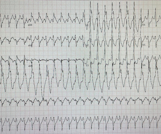
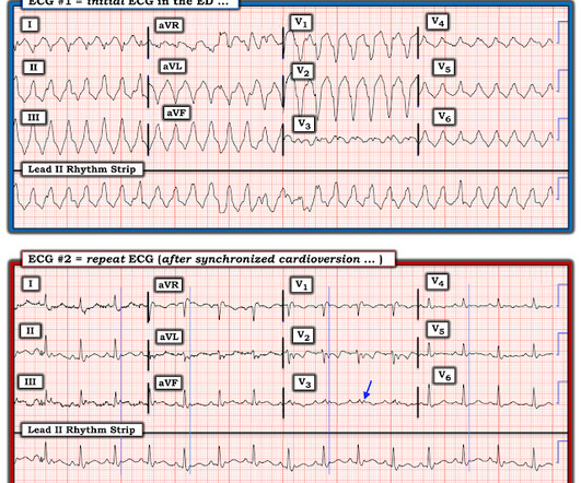
He denied any known history of CAD, but did report ASCVD risk factors to include HTN, HLD, and DM. Readers of the Smith ECG Blog will probably recognize this a very subtle inferior OMI. The VT vs SVT with Aberrancy debate is beyond the scope of this particular blog post. Here is the ECG after 200J. Examples provided below.
By Magnus Nossen This ECG is from a young man with no risk factors for CAD, he presented with chest pain. Before the lab values returned this patient had a n emergent coronary CT angiogram done that ruled out CAD. Each main coronary artery (LAD, RCA and LCx) are shown in separate images. There are no coronary stenoses.
This was sent by an undergraduate (not yet in medical school, but applying now) who works as an ED technician (records all EKGs, helps with procedures, takes vital signs) and who reads this blog regularly. They too have dense white masses consistent with coronary atherosclerosis. Edited by Smith He also sent me this great case.
The patient was treated as possible NSTEMI and underwent coronary angiography about 4 hours after presentation. TIMI 3 means the rate of passage of dye through the coronary artery is normal by angiography.) The electrophysiologist is a reader of Dr. Smith's ECG Blog. Initial hsTnI was 384 ng/L. He did not have access to ECG 1.
The ECG is just a test: a Bayesian approach to acute coronary occlusion If a patient with a recent femur fracture has sudden onset of pleuritic chest pain, shortness of breath, and hemoptysis, the D-dimer doesn’t matter: the patient’s pre-test likelihood for PE is so high that they need a CT. A Bayesian approach to acute coronary occlusion.
Angiogram: Severe two-vessel coronary artery disease with possible co-culprits (90% proximal circumflex, 70% mid/distal RCA) in the setting of non-ST elevation myocardial infarction. Marked ST depression from multi-vessel coronary disease serves to attentuate what would have been ST elevation in leads II and aVF ).
A man in his mid 60s with history of CAD and stents experienced sudden onset epigastric abdominal pain radiating up into his chest at home, waking him from sleep. This patient in today's case was a man in his 60s with a known history of coronary disease, including prior stents. This is a re-post of an excellent case from 2021.
A CT Coronary angiogram was ordered. Here are the results: --Minimally obstructive coronary artery disease. --LAD CAD-RADS category 1. --No Although a lesion is not visible anatomically on this CT scan, coronary catheter angiography could be considered based on Cardiology evaluation." A repeat troponin returned at 0.45
Post by Smith and Meyers Sam Ghali ( [link] ) just asked me (Smith): "Steve, do left main coronary artery *occlusions* (actual ones with transmural ischemia) have ST Depression or ST Elevation in aVR?" Furthermore, among 35 patients with acute left main coronary artery occlusion, 9 presented with RBBB (mostly with LAFB) on the admission ECG.
He has a history of known CAD, diabetes, and dyslipidemia. The ED ECG in the context of the prehospital ECGs was indeed diagnostic of acute coronary occlusion. Cath Results: The cath lab was activated and co-culprit lesions were found: 99% circumflex and 95% right coronary artery (RCA). Both were stented.
The biphasic T wave is consistent with recent reperfusion of an occluded coronary artery supplying the inferior region. Here’s the angiogram of the RCA : No thrombus or plaque rupture in the RCA (or any coronary artery) was found. This MI wasn’t caused by a ruptured plaque of CAD - it was a coronary artery dissection of the RCA.
This is for the version housed on Telegram: [link] You can get the full PM Cardio app here if you live in the UK or EU (or say you do upon registration): [link] Case Continued The cath lab was activated and the patient received 180 mg of ticagrelor, and then was transported to the cath lab.
Angiography showed normal coronaries. MINOCA: Myocardial Infarction in the Absence of Obstructive Coronary Artery Disease). Here is my comment on MINOCA: "Non-obstructive coronary disease" does not necessarily imply "no plaque rupture with thrombus." 2) overlooked obstructive coronary disease (e.g., The K was normal.
The ED provider ordered a coronary CT scan to assess the patient for CAD. The patient was taken emergently to the cath lab for a pericardiocentesis instead of a coronary angiogram. Three months prior to this presentation, he received a pacemaker for severe bradycardia and syncope due to sinus node dysfunction.
Hospital Course The patient was taken emergently to the cath lab which did not reveal any significant coronary artery disease, but she was noted to have reduced EF consistent with Takotsubo cardiomyopathy. Just because you don't see hemodynamically significant CAD on angiogram does not mean it is not OMI. It can only be seen by IVUS.
A man in his 70s with past medical history of hypertension, dyslipidemia, CAD s/p left circumflex stent 2 years prior presented to the ED with worsening intermittent exertional chest pain relieved by rest. The De Winter ECG pattern: morphology and accuracy for diagnosing acute coronary occlusion: systematic review. 2009;95:1701–1706.
Sent by Anonymous, written by Pendell Meyers A man in his 60s with history of CAD and 2 prior stents presented to the ED complaining of acute heavy substernal chest pain that began while eating breakfast about an hour ago, and had been persistent since then, despite EMS administering aspirin and nitroglycerin. Pre-intervention.
No family history of sudden cardiac death, cardiomyopathy, premature CAD, or other cardiac issues. Repeat CT angio chest (not CT coronary, unclear what protocol) showed possible LAD aneurysm and thrombus. Acute coronary occlusion almost always occurs in patients who are well beyond their teenage years.
The patient proceeded to cath where all coronaries were described as normal with no evidence of any CAD, spasm, or any other abnormality. She has not had a heart catheterization or after this event so the presence or absence of CAD is still unknown.
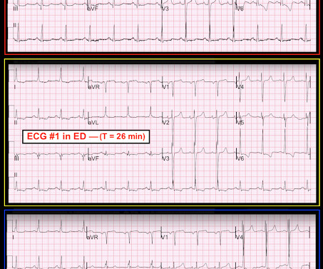
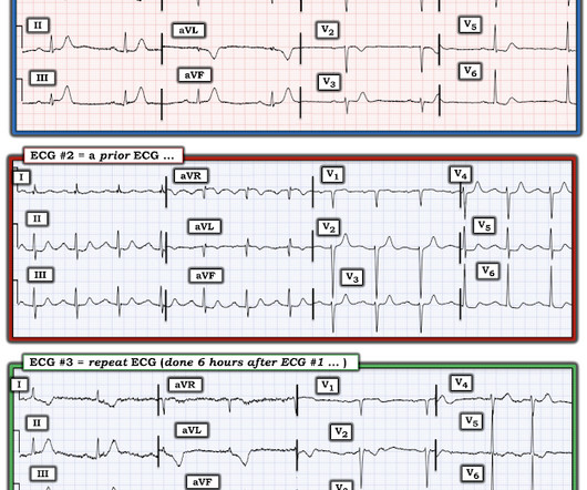
Concerning history, known CAD" Recorded 2 hours after pain onset: What do you think? To realize — Assessment of ECG #1 is complicated by knowing: i ) That today’s patient has a history of documented CAD ; and , ii ) The lack o f a prior tracing for comparison at the time the initial ECG was interpreted.
He had a history of CAD with CABG. Ventricular fibrillation is not only caused by acute coronary syndrome. A middle-aged male had a V Fib arrest. He had not complained of any premonitory symptoms (which is very common). Here was his initial ED ECG: There is atrial fibrillation with a rapid ventricular response. He did not have ACS.
But it does prove that the patient has coronary disease and makes the probability that his chest pain is due to ACS very very high. Instantaneous wave-free ratio is performed using high fidelity pressure wires that are passed distal to the coronary stenosis. Acute T-waves are large, even if not necessarily hyperacute.
She had zero CAD risk factors. Next day, t he patient was taken for an angiogram and found to have a reperfused LAD lesion with good flow that appeared to the angiographer as if it was a spontaneous coronary artery dissection. What is Spontaneous Coronary Artery Dissection (SCAD)? hours of substernal chest pressure.
I want all to know that, with the right mind preparation, and the use of the early repol/LAD occlusion formula, extremely subtle coronary occlusion can be detected prospectively, with no other information than the ECG. It is not a missed STEMI, but it is a missed coronary occlusion. Wang T, Zhang M, Fu Y, et al.
As in all ischemia interpretations with OMI findings, the findings can be due to type 1 AMI (example: acute coronary plaque rupture and thrombosis) or type 2 AMI (with or without fixed CAD, with severe regional supply/demand mismatch essentially equaling zero blood flow).
A middle aged male with no h/o CAD presented with one week of crescendo exertional angina, and had chest pain at the time of the first ECG: Here is the patient's previous ECG: Here is the patient's presenting ED ECG: There is isolated ST depression in precordial leads, deeper in V2 - V4 than in V5 or V6. There is no ST elevation.
He had a family history of early CAD and occasional drug and tobacco use. However, subtle coronary occlusion may be completely missed by the computer and called "normal." It is not yet available, but this is your way to get on the list. link] Here is the history: A 30 yo man presented complaining of severe chest pain. References : 1.
Is this due to coronary occlusion? The medic activated the cath lab but was refused by the interventionalist, who did not believe that this ECG represented acute coronary occlusion. But what we truly care about is coronary occlusion, for which STEMI is just a surrogate that is only about 75% sensitive for occlusion.
Submitted and written by Alex Bracey with edits by Pendell Meyers and Steve Smith Case A 50ish year old man with a history of CAD w/ prior LAD MI s/p LAD stenting presented to the ED with chest pain similar to his prior MI, but worse. Despite having acute coronary occlusion by cath, his ECGs never met STEMI criteria.
They found non-obstructive CAD, with only a 20% stenosis of OM2 and 10% RCA. As we have described multiple times on this blog, false positive "pericarditis" kills by distracting the clinician from actual emergencies including OMI, dissection, PE, and others. A repeat ECG was performed and cardiology was re-consulted: Roughly unchanged.
Late Gadolinium enhancement: Multifocal scarring of the septum (including RV septum), basal anterior wall and transmural mid inferior region scarring - a non-CAD hyperenhacement pattern. For review of a case of RVOT VT — Please see My Comment at the bottom of the page in the February 14, 2022 post in Dr. Smith's ECG Blog.
A middle-aged male with h/o CAD and stents presented with typical chest pressure. Is there likely to be fixed coronary stenosis that led to demand ischemia during pneumonia? --Was Furthermore, among 35 patients with acute left main coronary artery occlusion, 9 presented with RBBB (mostly with LAFB) on the admission ECG.
The diagnostic coronary angiogram identified only minimal coronary artery disease, but there was a severely calcified, ‘immobile’ aortic valve. Author continued : STE in aVR is often due to left main coronary artery obstruction (OR 4.72), and is associated with in-hospital cardiovascular mortality (OR 5.58).
The patient was transferred immediately for angiogram which revealed no significant CAD, and no intervention was performed. Coronary spasm causing massive current of injury with shark fin ECG. I would not expect ST-E to vanish in four beats with dissolving thrombus (also we know that the coronaries were clean).
A 75 yo with h/o CAD, CABG, and HFrEF presented after a syncopal episode. Discussion Thus, no further ECGs were recorded and there was no angiogram or stress test or CT coronary angiogram. There was no prodrome and no associated symptoms such as SOB or CP. The medics were worried about STEMI, as it meets STEMI criteria.
These findings are very subtle but suspicious for LAD occlusion, as we have seen in many similar (but less difficult) cases on this blog: A man in his sixties with chest pain at midnight with undetectable troponin How long would you like to wait for your Occlusion MI to show a STEMI? He also had non-acute CAD of the RCA (50%) and LCX (50%).
Case history A middle-aged woman with a history of HTN, but no prior CAD, presented to the ED with chest pain. LVH can mimic an acute anterior coronary occlusion (ACO) on the ECG. Electrocardiographic left ventricular hypertrophy in chest pain patients: Differentiation from acute coronary ischemic events. J Electrocardiol.
Second , the increased demand created by extreme tachycardia may exceed the ability of the coronary arteries to supply sufficient blood (due to preexisting three vessel or left main disease, with or without ACS). The lower heart rate was maintained, as were the ST segment changes above, over the next 10 minutes in the resuscitation room.
In this study of dialysis patients with severe CAD, 77% had an abnormal resting EKG and the most common abnormality was LVH. Another reason may be that the EKG is more difficult to interpret in patients with dialysis due to baseline abnormalities, including LVH. Herzog et al.
She also had non-acute CAD of the left main (50%) and LCX (75%). The Portable Programmable Microprocessor-Driven Real-Time 12-Lead Electrocardiographic Monitor: A Preliminary Report of a New Device for the Noninvasive Detection of Successful Reperfusion or Silent Coronary Reocclusion. They opened it. Initial troponin T was 0.46
He had significant history of CAD with CABG x5, and repeat CABG x 2 as well as a subsequent PCI of the graft to the RCA (twice) and of the graft to the Diagonal. Over a 13-month period, serum potassium and magnesium levels were measured in 590 patients admitted to a coronary care unit. Most recent echo showed EF of 60%.
Written by Jesse McLaren, with comments from Smith An 85 year old with a history of CAD presented with 3 hours of chest pain that feels like heartburn but that radiates to the left arm. Use STEMI criteria to identify acute coronary occlusion: the ECG was STEMI negative 2. Below is the ECG. What do you think?
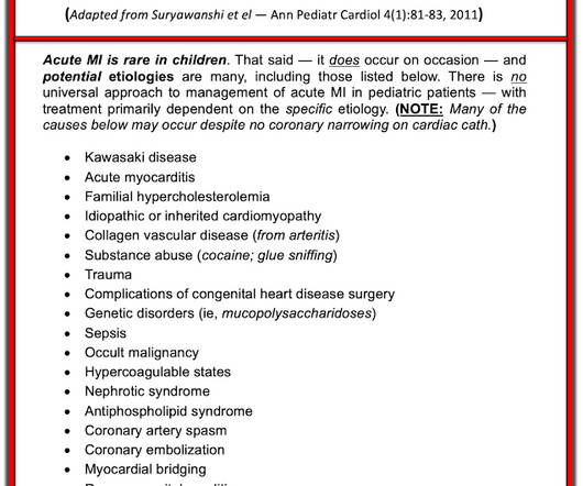
Acute coronary syndrome in a pediatric patient? He did have a family history notable for early CAD. hematological disorder like sickle cell or antiphospholipid syndome, family history of CAD or hypercholesterolemia, prior history of vasculopathies such as Kawasaki Disease, MIS-C, prior cardiac surgery, etc.)
Written by Pendell Meyers A woman in her 70s with known prior coronary artery disease experienced acute chest pain and shortness of breath. Her history and ECG were interpreted as very concerning for acute coronary syndrome which might benefit from acute reperfusion therapy. Vital signs were within normal limits. hours since onset.
We organize all of the trending information in your field so you don't have to. Join 5,000+ users and stay up to date on the latest articles your peers are reading.
You know about us, now we want to get to know you!
Let's personalize your content
Let's get even more personalized
We recognize your account from another site in our network, please click 'Send Email' below to continue with verifying your account and setting a password.


Let's personalize your content