This site uses cookies to improve your experience. To help us insure we adhere to various privacy regulations, please select your country/region of residence. If you do not select a country, we will assume you are from the United States. Select your Cookie Settings or view our Privacy Policy and Terms of Use.
Cookie Settings
Cookies and similar technologies are used on this website for proper function of the website, for tracking performance analytics and for marketing purposes. We and some of our third-party providers may use cookie data for various purposes. Please review the cookie settings below and choose your preference.
Used for the proper function of the website
Used for monitoring website traffic and interactions
Cookie Settings
Cookies and similar technologies are used on this website for proper function of the website, for tracking performance analytics and for marketing purposes. We and some of our third-party providers may use cookie data for various purposes. Please review the cookie settings below and choose your preference.
Strictly Necessary: Used for the proper function of the website
Performance/Analytics: Used for monitoring website traffic and interactions
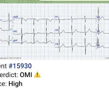
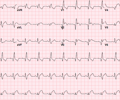
David Didlake EMT-P, RN, ACNP @DidlakeDW An adult male self-presented to the ED with palpitations and the following ECG. He denied any known history of CAD, but did report ASCVD risk factors to include HTN, HLD, and DM. Readers of the Smith ECG Blog will probably recognize this a very subtle inferior OMI.
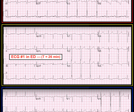
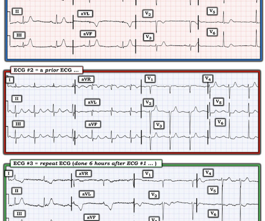
While in the ED, patient developed acute dyspnea while at rest, initially not associated with chest pain. The patient had no chest symptoms until he had been in the ED for many hours and had been undergoing management of his DKA. The patient was under the care of another ED physician. Another ECG was recorded: What do you think?
This was sent by an undergraduate (not yet in medical school, but applying now) who works as an ED technician (records all EKGs, helps with procedures, takes vital signs) and who reads this blog regularly. The patient re-presented to the ED a few days after his discharge with syncope.
Cardiology refused to be the admitting physician because it was "NSTEMI", and forced the ED physician to admit the patient to the hospitalist. Of course, there was terrible boarding and the patient was considered non-emergent (NSTEMI), and so could not leave the ED for some time. Scattered other nonobstructive CAD.
The patient presented to an outside hospital An 80yo female per triage “patient presents with chest pain, also hurts to breathe” PMH: CAD, s/p stent placement, CHF, atrial fibrillation, pacemaker (placed 1 month earlier), LBBB. This case was sent by Amandeep (Deep) Singh at Highland Hospital, part of Alameda Health System.
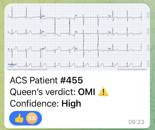
Here is what the Queen of Hearts AI app says: The patient received aspirin and NTG prehospital, and was transported to the ED. It could be a proximal RCA with both inferior OMI, posterior OMI (pulling ST down in V1/V2), and RV OMI causing large ischemic T-waves in V3-4.
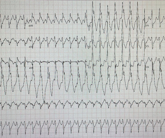
Despite otherwise normal vital signs, she was appropriately triaged to the critical care area of the ED. They are rare and hard to find in normal practice in the ED. For review of a case of RVOT VT — Please see My Comment at the bottom of the page in the February 14, 2022 post in Dr. Smith's ECG Blog. RVEF 100 ml/m2.
A man in his mid 60s with history of CAD and stents experienced sudden onset epigastric abdominal pain radiating up into his chest at home, waking him from sleep. He called EMS who brought him to the ED. ED Diagnoses: 1. We've come a long way in 2 years! And the pace only quickens. I ordered morphine but he refused.
Their feedback represent ed over 957 incidents overall and provided a ton of information to help iron out some of the initial wrinkles. With API , participating CAD and RMS vendors will be able to automatically send data back and forth to NERIS.
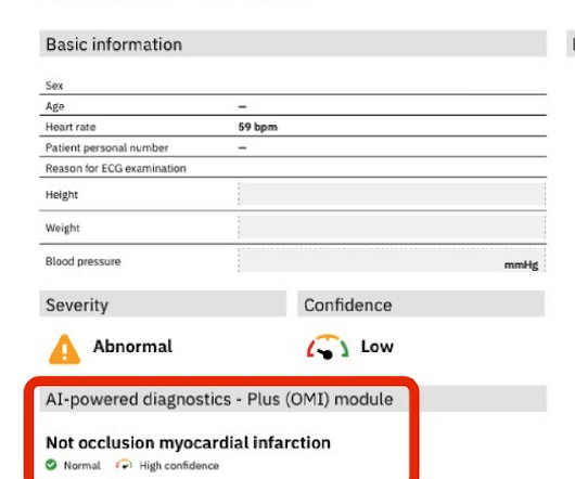
A middle-aged man complained of 15 minutes of classic angina that resolved upon arrival to the ED. So I made an ED diagnosis of Non-Occlusion Myocardial Infarction (NOMI), and his next day angiogram confirmed NOMI. Figure-1: The initial ECG that was done in the ED ( See text ). Here is his initial ECG: What do you think?
Case An 82 year old man with a history of hypertension presented to the ED with chest pain at 1211. The ED provider ordered a coronary CT scan to assess the patient for CAD. His pain suddenly became much worse in the ED and he became acutely diaphoretic, dizzy, and hypotensive. Another blood pressure was checked.
Sent by Anonymous, written by Pendell Meyers A man in his 60s with history of CAD and 2 prior stents presented to the ED complaining of acute heavy substernal chest pain that began while eating breakfast about an hour ago, and had been persistent since then, despite EMS administering aspirin and nitroglycerin. Pre-intervention.
An ECG was performed in the ED at 1554: Original image unavailable, this is the only recorded scanned ECG available. QOH Interpretation: The initial troponin I (older generation) at the first ED was barely positive at 0.06 She has not had a heart catheterization or after this event so the presence or absence of CAD is still unknown.
link] A 30 year-old woman was brought to the ED with chest pain. However, a smooth tapering of the mid-RCA was seen, highlighted in red below: How do we explain the MI if no sign of CAD was found? This MI wasn’t caused by a ruptured plaque of CAD - it was a coronary artery dissection of the RCA. This is written by Brooks Walsh.
This was a middle aged female with a h/o CAD who presented to the ED by EMS sudden onset of central chest pressure 45 min prior to ED arrival with associated diaphoresis and SOB. There is LVH and there are ST-T abnormalities (large inferior T-waves and ST elevation, with reciprocal findings in aVL).
He had a history of CAD with CABG. Here was his initial ED ECG: There is atrial fibrillation with a rapid ventricular response. A middle-aged male had a V Fib arrest. He had not complained of any premonitory symptoms (which is very common). There is profound ST depression especially in I, II, V2-V6.
So, I'm a follower of your blog, and I think I have a interesting case that I attended yesterday." Case "Male, 43yo, come to ED with Epigastric Pain started 3 hours ago. Remember: these findings above are included as STEMI equivalent findings in the 2022 ACC Expert Consensus Decision Pathway on ACS Patients in the ED.
Diagnosis of MINOCA should be made according to the Fourth Universal Definition of MI, in the absence of obstructive coronary artery disease (CAD) (no lesion ≥50%). The authors recommend using optical coherence tomography or intravascular ultrasound imaging in patients with evidence of nonobstructive CAD by angiogram. myocarditis).
A man in his 70s with past medical history of hypertension, dyslipidemia, CAD s/p left circumflex stent 2 years prior presented to the ED with worsening intermittent exertional chest pain relieved by rest. Written by Nathanael Franks MD, reviewed by Meyers, Smith, Grauer, etc.
He reportedly told his family "I think I'm having a heart attack", then they immediately drove him to the ED, and he was able to ambulate into the triage area before he collapsed and became unresponsive. The value of Stat Echo in the ED for confirming clinical and ECG suspicion of acute PE cannot be overstated!
A formal echocardiogram was completed the next day and again showed a normal ejection fraction without any focal wall motion abnormalities to suggest CAD. She has not had a heart catheterization or after this event so the presence or absence of CAD is still unknown. The Troponin I was cycled over time and was 0.353 followed by 0.296.
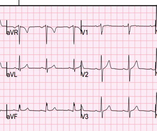
Concerning history, known CAD" Recorded 2 hours after pain onset: What do you think? To realize — Assessment of ECG #1 is complicated by knowing: i ) That today’s patient has a history of documented CAD ; and , ii ) The lack o f a prior tracing for comparison at the time the initial ECG was interpreted.
They found non-obstructive CAD, with only a 20% stenosis of OM2 and 10% RCA. As we have described multiple times on this blog, false positive "pericarditis" kills by distracting the clinician from actual emergencies including OMI, dissection, PE, and others. A repeat ECG was performed and cardiology was re-consulted: Roughly unchanged.
A man is his late 50’s presents to the ED with 1 hour of post exertional chest pressure associated with diaphoresis and nausea. He has a history of known CAD, diabetes, and dyslipidemia. The ED ECG in the context of the prehospital ECGs was indeed diagnostic of acute coronary occlusion. Leads II, III, aVF show about 0.5
He had a family history of early CAD and occasional drug and tobacco use. The ECG was alarming to the ED physician who did indeed review it. It is not yet available, but this is your way to get on the list. link] Here is the history: A 30 yo man presented complaining of severe chest pain.
These findings are very subtle but suspicious for LAD occlusion, as we have seen in many similar (but less difficult) cases on this blog: A man in his sixties with chest pain at midnight with undetectable troponin How long would you like to wait for your Occlusion MI to show a STEMI? He also had non-acute CAD of the RCA (50%) and LCX (50%).
CAD-RADS category 1. --No That said — I did not feel the history we were given pointed to any particular diagnosis ( ie, 3 episodes of CP and dyspnea of uncertain duration over the past day — with pain on deep breathing — but with symptoms apparently resolved by the time the patient arrived in the ED ).
Submitted and written by Alex Bracey with edits by Pendell Meyers and Steve Smith Case A 50ish year old man with a history of CAD w/ prior LAD MI s/p LAD stenting presented to the ED with chest pain similar to his prior MI, but worse. Around 19 hours later, he experienced the same pain, which prompted his presentation to the ED.
Here is a case that demonstrates this very well: Isolated "Inferior" ST Segment Depression: Not a Sign of Inferior Ischemia Here is the most viewed post of all time on Dr. Smith's ECG Blog, with nearly 100,000 views: Five Primary Patterns of Ischemic ST depression, without ST elevation. Use ED Echo if available 4. Look at aVF.
A 75 yo with h/o CAD, CABG, and HFrEF presented after a syncopal episode. Of interest Lead I on the initial ECG from the ED = ECG #2 ( which was done a bit after the prehospital ECG #1 was done ) no longer shows this unusual 4-phase QRS deflection in lead I. There was no prodrome and no associated symptoms such as SOB or CP.
J Electrocardiol [Internet] 2022;Available from: [link] Cardiology opinion: Takotsubo Cardiomyopathy (EF 30-35%) V Fib Cardiac arrest Prolonged QTC NSTEMI (Smith comment: is it NSTEMI or is it Takotsubo? -- these are entirely different) Moderate single-vessel CAD. I could have told you this (and did tell you this) without an MRI.
A middle-aged male with h/o CAD and stents presented with typical chest pressure. The patient arrived in the ED and had this ECG recorded: Interpretation? Here is his ECG: The resident was alarmed at the "ST elevation in III with reciprocal ST depression in aVL" Are you alarmed? This is a very common misread.
A middle aged male with no h/o CAD presented with one week of crescendo exertional angina, and had chest pain at the time of the first ECG: Here is the patient's previous ECG: Here is the patient's presenting ED ECG: There is isolated ST depression in precordial leads, deeper in V2 - V4 than in V5 or V6. There is no ST elevation.
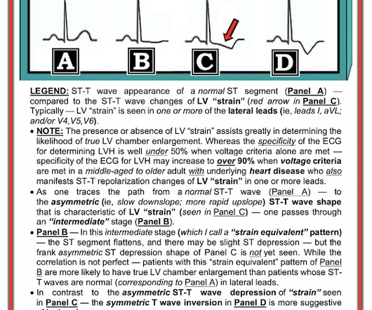
This was my thought: if this patient presented to the ED with chest pain, then this is an LAD occlusion. See image below: Slow upstroke, fast downstroke. Asymmetric.
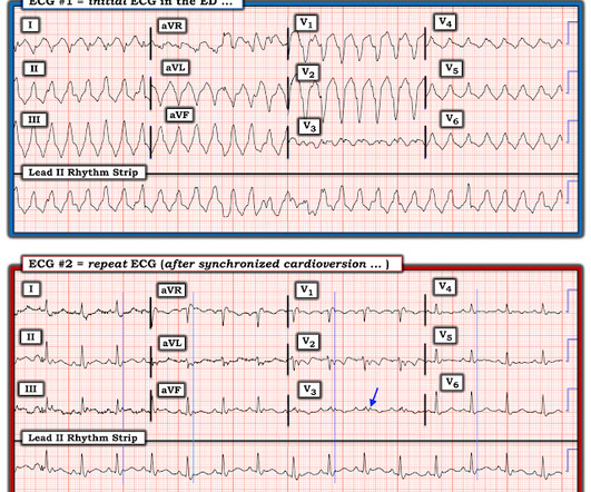
The case An older woman presented to the ED with dyspnea, diaphoresis, and chest pressure. She had a normal EF, and no significant CAD, and was taking flecainide to suppress the AF. Figure-1: TOP — ECG #1 ( = the initial ECG performed in the ED). The initial ECG: The rhythm is regular at 150, and the QRS is markedly wide.
No family history of sudden cardiac death, cardiomyopathy, premature CAD, or other cardiac issues. No similar symptoms in the past. No prior exertional complaints of chest pain, dizziness, lightheadedness, or undue shortness of breath. He denied headache or neck pain associated with exertion.
His ED cardiac ultrasound (which is not at all ideal for detecting wall motion abnormalities, and is also very operator dependent for this finding) was significant for depressed global EF. In this study of dialysis patients with severe CAD, 77% had an abnormal resting EKG and the most common abnormality was LVH. Herzog et al.
Smith and Meyers answer: First , LM occlusion is uncommon in the ED because most of these die before they can get a 12-lead recorded. Beware crescendo angina in patient with known CAD ST Elevation in aVR Case 7. TIMI flow 0) is rare in the ED, as most either die before arrival or are recognized clinically due to cardiogenic shock.
She had zero CAD risk factors. The 1st “lesson” is, “All bets are off” — when an adult of any age presents to the ED with new-onset chest discomfort. hours of substernal chest pressure. It was non-radiating and without other associated symptoms except for nausea. Here was her ECG at time zero: What do you think?
She was asymptomatic at the time of this ECG recorded on arrival to our ED: What do you think? She also had non-acute CAD of the left main (50%) and LCX (75%). By the time the patient arrived at our facility, she had received aspirin and nitroglycerin, and her pain had apparently completely resolved. They opened it.
Case history A middle-aged woman with a history of HTN, but no prior CAD, presented to the ED with chest pain. There is ST elevation, but also high voltage (though the high voltage is NOT in the leads with worrisome STE, rather, it is in aVL). Is the ST elevation due to LVH? Her vitals signs were remarkable for marked hypertension.
The patient was brought directly to the cardiac catheterization lab for PCI, bypassing the ED. As I met the paramedics and cath team in the lab, I was ready to see severe coronary disease (CAD), but the vessels were non-obstructive. Folland ED, et al. In the cath lab, the patient’s blood pressure remained low. De Backer D et al.
He had significant history of CAD with CABG x5, and repeat CABG x 2 as well as a subsequent PCI of the graft to the RCA (twice) and of the graft to the Diagonal. Here is his ED ECG: There is obvious infero-posterior STEMI. A late middle-aged man presented with one hour of chest pain. Most recent echo showed EF of 60%.
Written by Jesse McLaren, with comments from Smith An 85 year old with a history of CAD presented with 3 hours of chest pain that feels like heartburn but that radiates to the left arm. The Repeat ECG: As per Dr. McLaren — the patient was unfortunately discharged from the ED — but returned 6 hours later with a recurrence of chest pain.
We organize all of the trending information in your field so you don't have to. Join 5,000+ users and stay up to date on the latest articles your peers are reading.
You know about us, now we want to get to know you!
Let's personalize your content
Let's get even more personalized
We recognize your account from another site in our network, please click 'Send Email' below to continue with verifying your account and setting a password.


Let's personalize your content