This site uses cookies to improve your experience. To help us insure we adhere to various privacy regulations, please select your country/region of residence. If you do not select a country, we will assume you are from the United States. Select your Cookie Settings or view our Privacy Policy and Terms of Use.
Cookie Settings
Cookies and similar technologies are used on this website for proper function of the website, for tracking performance analytics and for marketing purposes. We and some of our third-party providers may use cookie data for various purposes. Please review the cookie settings below and choose your preference.
Used for the proper function of the website
Used for monitoring website traffic and interactions
Cookie Settings
Cookies and similar technologies are used on this website for proper function of the website, for tracking performance analytics and for marketing purposes. We and some of our third-party providers may use cookie data for various purposes. Please review the cookie settings below and choose your preference.
Strictly Necessary: Used for the proper function of the website
Performance/Analytics: Used for monitoring website traffic and interactions
At the time of ED arrival he was alert, oriented, and verbalizing only a headache with a normalized BP. The ED activated trauma services, and a 12 Lead ECG was captured. This was deemed “non-specific” by the ED physicians. Thus, the ED admission ECG changes cannot be blamed on LVH. The fall was not a mechanical etiology.
Major adverse cardiac event rates in moderate-risk patients: Does prior coronary disease matter? Major adverse cardiac event rates in moderate-risk patients: Does prior coronary disease matter? Case: You are working a shift in your local community emergency department (ED) when a 47-year-old male presents with chest pain.
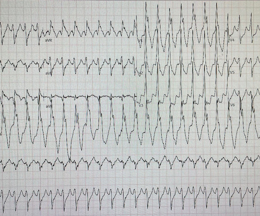
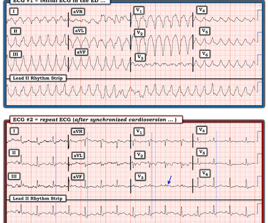
David Didlake EMT-P, RN, ACNP @DidlakeDW An adult male self-presented to the ED with palpitations and the following ECG. He denied any known history of CAD, but did report ASCVD risk factors to include HTN, HLD, and DM. The patient was very uncomfortable, dyspneic, and displayed an SpO2 90% on RA.
While in the ED, patient developed acute dyspnea while at rest, initially not associated with chest pain. The patient had no chest symptoms until he had been in the ED for many hours and had been undergoing management of his DKA. The patient was under the care of another ED physician. Another ECG was recorded: What do you think?
This was sent by an undergraduate (not yet in medical school, but applying now) who works as an ED technician (records all EKGs, helps with procedures, takes vital signs) and who reads this blog regularly. They too have dense white masses consistent with coronary atherosclerosis. Edited by Smith He also sent me this great case.
David Didlake Acute Care Nurse Practitioner Firefighter / Paramedic (ret) @DidlakeDW Expert commentary and peer review by Dr. Steve Smith [link] @smithECGBlog A 57 y/o Female with PMHx HTN, HLD, DM, and current use of tobacco products, presented to the ED with chest discomfort. It’s judicious, then, to arrange for coronary angiogram.
Moreover, he had no pertinent medical history to report in terms of CAD, HTN, HLD, or DM, for example. One cannot rely on this feature as a means of detecting changes – subtle, or dramatic – for volatile occlusive coronary thrombus. Here is the final ECG just prior to ED transfer. A 12 Lead ECG was recorded.
A man in his mid 60s with history of CAD and stents experienced sudden onset epigastric abdominal pain radiating up into his chest at home, waking him from sleep. He called EMS who brought him to the ED. ED Diagnoses: 1. We've come a long way in 2 years! And the pace only quickens. I ordered morphine but he refused.
Here is what the Queen of Hearts AI app says: The patient received aspirin and NTG prehospital, and was transported to the ED. It could be a proximal RCA with both inferior OMI, posterior OMI (pulling ST down in V1/V2), and RV OMI causing large ischemic T-waves in V3-4.
A man is his late 50’s presents to the ED with 1 hour of post exertional chest pressure associated with diaphoresis and nausea. He has a history of known CAD, diabetes, and dyslipidemia. The ED ECG in the context of the prehospital ECGs was indeed diagnostic of acute coronary occlusion. Leads II, III, aVF show about 0.5
A CT Coronary angiogram was ordered. Here are the results: --Minimally obstructive coronary artery disease. --LAD CAD-RADS category 1. --No Although a lesion is not visible anatomically on this CT scan, coronary catheter angiography could be considered based on Cardiology evaluation." A repeat troponin returned at 0.45
link] A 30 year-old woman was brought to the ED with chest pain. The biphasic T wave is consistent with recent reperfusion of an occluded coronary artery supplying the inferior region. Here’s the angiogram of the RCA : No thrombus or plaque rupture in the RCA (or any coronary artery) was found. This is written by Brooks Walsh.
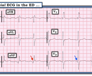
A middle-aged man complained of 15 minutes of classic angina that resolved upon arrival to the ED. But it does prove that the patient has coronary disease and makes the probability that his chest pain is due to ACS very very high. Figure-1: The initial ECG that was done in the ED ( See text ).
Angiography showed normal coronaries. MINOCA: Myocardial Infarction in the Absence of Obstructive Coronary Artery Disease). Here is my comment on MINOCA: "Non-obstructive coronary disease" does not necessarily imply "no plaque rupture with thrombus." 2) overlooked obstructive coronary disease (e.g., The K was normal.
He had a history of CAD with CABG. Here was his initial ED ECG: There is atrial fibrillation with a rapid ventricular response. Ventricular fibrillation is not only caused by acute coronary syndrome. A middle-aged male had a V Fib arrest. He had not complained of any premonitory symptoms (which is very common).
A 59-year-old male with a past medical history of a repaired ventricular septal defect (VSD), dextrocardia, hypertension, hyperlipidemia, and current smoker presented to the emergency department (ED). This patient had known coronary artery disease (CAD), and previously required drug eluting stents to the obtuse marginal and diagonal arteries.
Case An 82 year old man with a history of hypertension presented to the ED with chest pain at 1211. The ED provider ordered a coronary CT scan to assess the patient for CAD. His pain suddenly became much worse in the ED and he became acutely diaphoretic, dizzy, and hypotensive. Another blood pressure was checked.
Sent by Anonymous, written by Pendell Meyers A man in his 60s with history of CAD and 2 prior stents presented to the ED complaining of acute heavy substernal chest pain that began while eating breakfast about an hour ago, and had been persistent since then, despite EMS administering aspirin and nitroglycerin. Pre-intervention.
She had zero CAD risk factors. Next day, t he patient was taken for an angiogram and found to have a reperfused LAD lesion with good flow that appeared to the angiographer as if it was a spontaneous coronary artery dissection. What is Spontaneous Coronary Artery Dissection (SCAD)? hours of substernal chest pressure.
A man in his 70s with past medical history of hypertension, dyslipidemia, CAD s/p left circumflex stent 2 years prior presented to the ED with worsening intermittent exertional chest pain relieved by rest. The De Winter ECG pattern: morphology and accuracy for diagnosing acute coronary occlusion: systematic review. As per Drs.
Hospital Course The patient was taken emergently to the cath lab which did not reveal any significant coronary artery disease, but she was noted to have reduced EF consistent with Takotsubo cardiomyopathy. Just because you don't see hemodynamically significant CAD on angiogram does not mean it is not OMI. It can only be seen by IVUS.
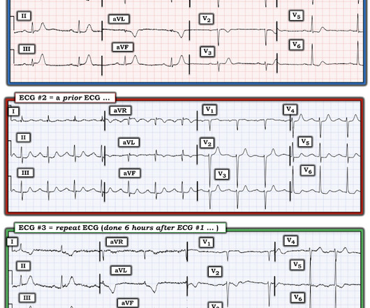
Concerning history, known CAD" Recorded 2 hours after pain onset: What do you think? To realize — Assessment of ECG #1 is complicated by knowing: i ) That today’s patient has a history of documented CAD ; and , ii ) The lack o f a prior tracing for comparison at the time the initial ECG was interpreted. What Do We Learn from ECG #3 ?
I want all to know that, with the right mind preparation, and the use of the early repol/LAD occlusion formula, extremely subtle coronary occlusion can be detected prospectively, with no other information than the ECG. This was my thought: if this patient presented to the ED with chest pain, then this is an LAD occlusion. Asymmetric.
He had a family history of early CAD and occasional drug and tobacco use. The ECG was alarming to the ED physician who did indeed review it. However, subtle coronary occlusion may be completely missed by the computer and called "normal." It is not yet available, but this is your way to get on the list. References : 1.
A middle aged male with no h/o CAD presented with one week of crescendo exertional angina, and had chest pain at the time of the first ECG: Here is the patient's previous ECG: Here is the patient's presenting ED ECG: There is isolated ST depression in precordial leads, deeper in V2 - V4 than in V5 or V6. There is no ST elevation.
Is this due to coronary occlusion? The medic activated the cath lab but was refused by the interventionalist, who did not believe that this ECG represented acute coronary occlusion. But what we truly care about is coronary occlusion, for which STEMI is just a surrogate that is only about 75% sensitive for occlusion. Look at aVF.
The patient was brought directly to the cardiac catheterization lab for PCI, bypassing the ED. The diagnostic coronary angiogram identified only minimal coronary artery disease, but there was a severely calcified, ‘immobile’ aortic valve. In the cath lab, the patient’s blood pressure remained low.
They found non-obstructive CAD, with only a 20% stenosis of OM2 and 10% RCA. IF , on the other hand — the patient with new chest pain is older and lacks predisposing viral symptoms — then acute pericarditis becomes a rare diagnosis in an ED setting. A repeat ECG was performed and cardiology was re-consulted: Roughly unchanged.
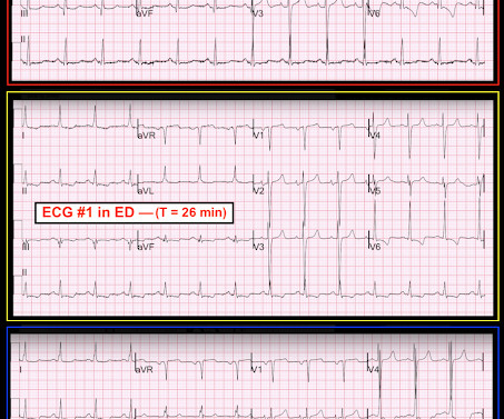
Cardiology was called and the patient was taken for urgent catheterization with the time from ED arrival to cath about 1 hour and 45 minutes. He also had non-acute CAD of the RCA (50%) and LCX (50%). I focus my att ention on the interpretation of the initial ED tracing ( = E CG # 1 in Figure-1 ).
Submitted and written by Alex Bracey with edits by Pendell Meyers and Steve Smith Case A 50ish year old man with a history of CAD w/ prior LAD MI s/p LAD stenting presented to the ED with chest pain similar to his prior MI, but worse. Around 19 hours later, he experienced the same pain, which prompted his presentation to the ED.
Case history A middle-aged woman with a history of HTN, but no prior CAD, presented to the ED with chest pain. LVH can mimic an acute anterior coronary occlusion (ACO) on the ECG. Electrocardiographic left ventricular hypertrophy in chest pain patients: Differentiation from acute coronary ischemic events. 2014.06.001.
A 75 yo with h/o CAD, CABG, and HFrEF presented after a syncopal episode. Discussion Thus, no further ECGs were recorded and there was no angiogram or stress test or CT coronary angiogram. There was no prodrome and no associated symptoms such as SOB or CP. The medics were worried about STEMI, as it meets STEMI criteria.
Despite otherwise normal vital signs, she was appropriately triaged to the critical care area of the ED. They are rare and hard to find in normal practice in the ED. She denied chest pain and denied feeling any palpitations, even during her triage ECG: What do you think? There is mild-moderate tricuspid valve regurgitation.
Furthermore, there was no family history of early CAD, MI, or sudden cardiac death. 1] Here is the admitting ED ECG after cancellation of Code STEMI. The patient continued to verbalize cessation of symptoms while in the ED. Cardiology admitted him for observation with plans for next-day coronary angiogram. 1] Driver, B.
Otherwise, no admission of CAD, HLD, or family history of sudden cardiac death. A second 12 Lead ECG was recorded: This is a testament to the dynamic nature of coronary thrombosis and thrombolysis. He described the pain as “nagging,” and equally not exacerbated by any kind of movement. But the lesion is still active!
Post by Smith and Meyers Sam Ghali ( [link] ) just asked me (Smith): "Steve, do left main coronary artery *occlusions* (actual ones with transmural ischemia) have ST Depression or ST Elevation in aVR?" Smith and Meyers answer: First , LM occlusion is uncommon in the ED because most of these die before they can get a 12-lead recorded.
Pervasive use of CT coronary angiography has been an unnecessary feature of the evaluation of patient with low-risk chest pain for the better part of a decade now. Patients were eligible by symptoms of an acute coronary syndrome, supported by ECG changes, an elevated troponin, or a history of ischemic heart disease.
An ECG was performed in the ED at 1554: Original image unavailable, this is the only recorded scanned ECG available. QOH Interpretation: The initial troponin I (older generation) at the first ED was barely positive at 0.06 She has not had a heart catheterization or after this event so the presence or absence of CAD is still unknown.
No family history of sudden cardiac death, cardiomyopathy, premature CAD, or other cardiac issues. Repeat CT angio chest (not CT coronary, unclear what protocol) showed possible LAD aneurysm and thrombus. Acute coronary occlusion almost always occurs in patients who are well beyond their teenage years.
He reportedly told his family "I think I'm having a heart attack", then they immediately drove him to the ED, and he was able to ambulate into the triage area before he collapsed and became unresponsive. The value of Stat Echo in the ED for confirming clinical and ECG suspicion of acute PE cannot be overstated!
A middle-aged male with h/o CAD and stents presented with typical chest pressure. The patient arrived in the ED and had this ECG recorded: Interpretation? Is there likely to be fixed coronary stenosis that led to demand ischemia during pneumonia? --Was This is a very common misread. The trick is to find the end of the QRS.
His ED cardiac ultrasound (which is not at all ideal for detecting wall motion abnormalities, and is also very operator dependent for this finding) was significant for depressed global EF. In this study of dialysis patients with severe CAD, 77% had an abnormal resting EKG and the most common abnormality was LVH. Herzog et al.
Written by Jesse McLaren, with comments from Smith An 85 year old with a history of CAD presented with 3 hours of chest pain that feels like heartburn but that radiates to the left arm. Use STEMI criteria to identify acute coronary occlusion: the ECG was STEMI negative 2. Below is the ECG. What do you think?
He had significant history of CAD with CABG x5, and repeat CABG x 2 as well as a subsequent PCI of the graft to the RCA (twice) and of the graft to the Diagonal. Here is his ED ECG: There is obvious infero-posterior STEMI. A late middle-aged man presented with one hour of chest pain. Most recent echo showed EF of 60%.
We organize all of the trending information in your field so you don't have to. Join 5,000+ users and stay up to date on the latest articles your peers are reading.
You know about us, now we want to get to know you!
Let's personalize your content
Let's get even more personalized
We recognize your account from another site in our network, please click 'Send Email' below to continue with verifying your account and setting a password.


Let's personalize your content