Remove CAD Remove Definition Remove ED
article thumbnail

Elder Male with Syncope

EMS 12-Lead

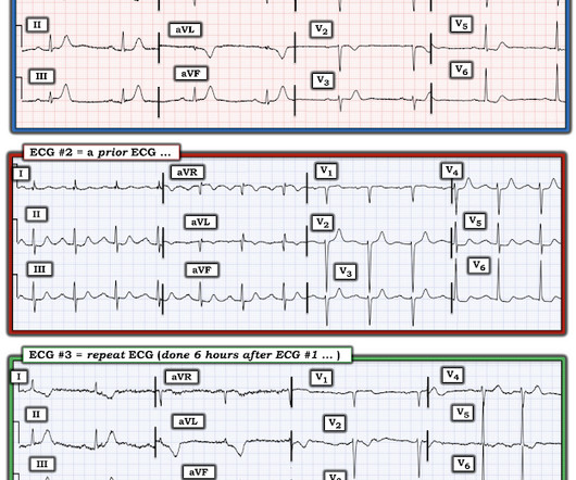
At the time of ED arrival he was alert, oriented, and verbalizing only a headache with a normalized BP. The ED activated trauma services, and a 12 Lead ECG was captured. This was deemed “non-specific” by the ED physicians. Thus, the ED admission ECG changes cannot be blamed on LVH. The fall was not a mechanical etiology.

Coronary 290
article thumbnail

Critical Left Main

EMS 12-Lead

David Didlake Acute Care Nurse Practitioner Firefighter / Paramedic (ret) @DidlakeDW Expert commentary and peer review by Dr. Steve Smith [link] @smithECGBlog A 57 y/o Female with PMHx HTN, HLD, DM, and current use of tobacco products, presented to the ED with chest discomfort. A 12 Lead ECG was captured on her arrival. Coronary Angiogram 1.

Coronary 130
professionals

Sign Up for our Newsletter

This site is protected by reCAPTCHA and the Google Privacy Policy and Terms of Service apply.

article thumbnail

1 hour of CPR, then ECMO circulation, then successful defibrillation.

Dr. Smith's ECG Blog

A followup ECG was recorded 2 days later: No definite evidence of infarction. Diagnosis of MINOCA should be made according to the Fourth Universal Definition of MI, in the absence of obstructive coronary artery disease (CAD) (no lesion ≥50%). For clarity — I’ll again show the initial ECG done in the ED in Figure-1.

article thumbnail

See this "NSTEMI" go unrecognized for what it really is, how it progresses, and what happens

Dr. Smith's ECG Blog

A man in his 70s with past medical history of hypertension, dyslipidemia, CAD s/p left circumflex stent 2 years prior presented to the ED with worsening intermittent exertional chest pain relieved by rest. Written by Nathanael Franks MD, reviewed by Meyers, Smith, Grauer, etc. Smith : I think it looks more like posterior OMI.

ACS 87
article thumbnail

Infection and DKA, then sudden dyspnea while in the ED

Dr. Smith's ECG Blog

While in the ED, patient developed acute dyspnea while at rest, initially not associated with chest pain. The patient had no chest symptoms until he had been in the ED for many hours and had been undergoing management of his DKA. The patient was under the care of another ED physician. Another ECG was recorded: What do you think?

ED 106
article thumbnail

A 50-Something Male with 2 hours of Chest discomfort

Dr. Smith's ECG Blog

Concerning history, known CAD" Recorded 2 hours after pain onset: What do you think? To realize — Assessment of ECG #1 is complicated by knowing: i ) That today’s patient has a history of documented CAD ; and , ii ) The lack o f a prior tracing for comparison at the time the initial ECG was interpreted.

STEMI 52
article thumbnail

Wide-complex tachycardia: VT, aberrant, or "other?"

Dr. Smith's ECG Blog

The case An older woman presented to the ED with dyspnea, diaphoresis, and chest pressure. She had a normal EF, and no significant CAD, and was taking flecainide to suppress the AF. Figure-1: TOP — ECG #1 ( = the initial ECG performed in the ED). The initial ECG: The rhythm is regular at 150, and the QRS is markedly wide.

OR 52