This site uses cookies to improve your experience. To help us insure we adhere to various privacy regulations, please select your country/region of residence. If you do not select a country, we will assume you are from the United States. Select your Cookie Settings or view our Privacy Policy and Terms of Use.
Cookie Settings
Cookies and similar technologies are used on this website for proper function of the website, for tracking performance analytics and for marketing purposes. We and some of our third-party providers may use cookie data for various purposes. Please review the cookie settings below and choose your preference.
Used for the proper function of the website
Used for monitoring website traffic and interactions
Cookie Settings
Cookies and similar technologies are used on this website for proper function of the website, for tracking performance analytics and for marketing purposes. We and some of our third-party providers may use cookie data for various purposes. Please review the cookie settings below and choose your preference.
Strictly Necessary: Used for the proper function of the website
Performance/Analytics: Used for monitoring website traffic and interactions
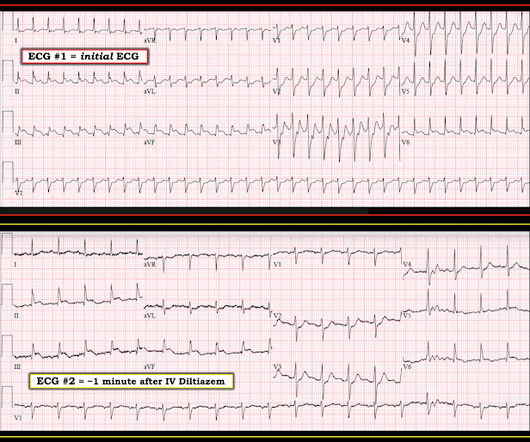
He had no previous history of CAD, and presented with very typical waxing and waning chest pain, much worse with exertion but also present at rest and on presentation, though his pain was minimal at the time of the ECG. This is all suggestive of posterior STEMI, but not definitely diagnostic. Blood pressure was 150/80.
20% of cases that everyone would call a STEMI have a competely open artery by the time of angiogram 60-90 minutes later. Patient stated that he has had glucose over 400 even though he has not missed any doses of insulin. Here is the ECG at that point in time: The ischemia is mostly resolved.
This is a troponin I level that is almost exclusively seen in STEMI. A followup ECG was recorded 2 days later: No definite evidence of infarction. So this is either a case of MINOCA, or a case of Type II STEMI. I believe the latter (type II STEMI) is most likely. pulmonary embolism, sepsis, etc.), myocarditis).
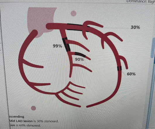
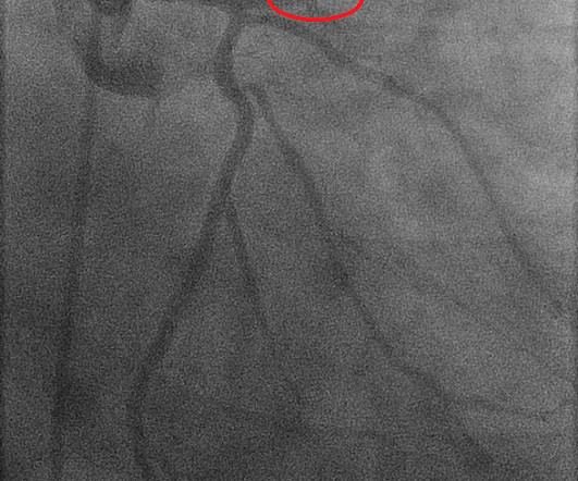
However, a smooth tapering of the mid-RCA was seen, highlighted in red below: How do we explain the MI if no sign of CAD was found? This MI wasn’t caused by a ruptured plaque of CAD - it was a coronary artery dissection of the RCA. A recent study found that SCAD causes almost 20% of STEMI in young women. A study by Hassan et al.
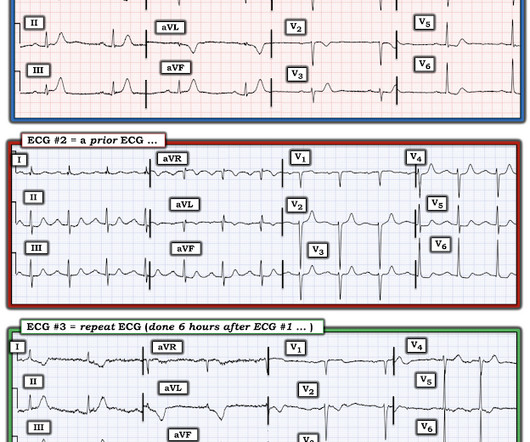
A prior ECG from 1 month ago was available: The presentation ECG was interpreted as STEMI and the patient was transferred emergently to the nearest PCI center. The patient proceeded to cath where all coronaries were described as normal with no evidence of any CAD, spasm, or any other abnormality. So maybe she is better than I am.
J Electrocardiol [Internet] 2022;Available from: [link] Cardiology opinion: Takotsubo Cardiomyopathy (EF 30-35%) V Fib Cardiac arrest Prolonged QTC NSTEMI (Smith comment: is it NSTEMI or is it Takotsubo? -- these are entirely different) Moderate single-vessel CAD. I could have told you this (and did tell you this) without an MRI.
Concerning history, known CAD" Recorded 2 hours after pain onset: What do you think? The patient was diagnosed with a"Non-STEMI." Traditionally , Occlusion MI (OMI) myocardial infarctions that are not STEMI are called NonSTEMI. This was my response: "This looks like a worrisome EKG. But by now you must have a repeat ECG.
CAD-RADS category 1. --No Later, she developed chest pain again, and had this ECG recorded: Obvious Anterior OMI that is also a STEMI Coronary angiogram- --Right dominant coronary artery system --The left main artery was normal in appearance and free of obstructive disease. --The Transient STEMI is at high risk of re-occlusion.
This is technically a STEMI, with 1.5 However, I think many practitioners might not see this as a clear STEMI, and would instead call this "borderline." They collected several repeat ECGs at the outside hospital before transport: None of these three ECGs meet STEMI criteria. This ECG was recorded on arrival: What do you think?
A man in his 70s with past medical history of hypertension, dyslipidemia, CAD s/p left circumflex stent 2 years prior presented to the ED with worsening intermittent exertional chest pain relieved by rest. Written by Nathanael Franks MD, reviewed by Meyers, Smith, Grauer, etc. In my opinion, I think it looks more like subendocardial ischemia.
Submitted and written by Alex Bracey with edits by Pendell Meyers and Steve Smith Case A 50ish year old man with a history of CAD w/ prior LAD MI s/p LAD stenting presented to the ED with chest pain similar to his prior MI, but worse. Lead aVL, for example, has a definite J-wave. Stat echo would also be helpful.
The axiom of "type 1 (ACS, plaque rupture) STEMIs are not tachycardic unless they are in cardiogenic shock" is not applicable outside of sinus rhythm. Is that an obvious STEMI underneath that rhythm? But this is not definite because the angiogram was not done at the same time as the ECG changes suggesting OMI.
A 75 yo with h/o CAD, CABG, and HFrEF presented after a syncopal episode. The medics were worried about STEMI, as it meets STEMI criteria. The troponins are NOT consistent with STEMI (OMI), which typically has a troponin I of at least 5 ng/mL. There was no prodrome and no associated symptoms such as SOB or CP.
These findings are very subtle but suspicious for LAD occlusion, as we have seen in many similar (but less difficult) cases on this blog: A man in his sixties with chest pain at midnight with undetectable troponin How long would you like to wait for your Occlusion MI to show a STEMI? He also had non-acute CAD of the RCA (50%) and LCX (50%).
2) The STE in V1 and V2 has an R'-wave and downsloping ST segments, very atypical for STEMI. Cardiology was consulted and they agreed that the EKG had an atypical morphology for STEMI and did not activate the cath lab. She has not had a heart catheterization or after this event so the presence or absence of CAD is still unknown.
Clinical Course The paramedic activated a “Code STEMI” alert and transported the patient nearly 50 miles to the closest tertiary medical center. 2 The astute paramedic recognized this possibility and announced a CODE STEMI. 6,7 Surgical repair of AS, by either TAVR or SAVR, is the definitive treatment for this condition.
Written by Jesse McLaren, with comments from Smith An 85 year old with a history of CAD presented with 3 hours of chest pain that feels like heartburn but that radiates to the left arm. There’s minimal concave ST elevation in III which does not meet STEMI criteria, so this ECG is "STEMI negative". Below is the ECG. Take home 1.
The procedure was described as very complex due to severe multivessel CAD, but ultimately PCI was successfully performed to the ostial LCX. Final Diagnosis: "STEMI" (of course, as you can see in the ECGs above, this is not true, by definition this was NSTEMI. Pre-intervention. In other words, millimeters really don't matter!
No prior similar symptoms or known CAD. The Queen of Hearts Diagnosed "STEMI/STEMI equivalent" on that first ECG (she now uses "STEMI Equivalent" rather than OMI). The fact that she states "STEMI-Equivalent" here means that she does not think it is reperfused, but she does not know that the patient is pain free now.
Written by Willy Frick A 52 year old man with hypertension, dyslipidemia, and seropositive rheumatoid arthritis (a risk factor for CAD) presented with acute substernal chest pressure with diaphoresis which woke him from sleep just after midnight. He said it felt like "someone ripped [his] heart out." J Cardiovascular Interventions.--20/53
We organize all of the trending information in your field so you don't have to. Join 5,000+ users and stay up to date on the latest articles your peers are reading.
You know about us, now we want to get to know you!
Let's personalize your content
Let's get even more personalized
We recognize your account from another site in our network, please click 'Send Email' below to continue with verifying your account and setting a password.


Let's personalize your content