This site uses cookies to improve your experience. To help us insure we adhere to various privacy regulations, please select your country/region of residence. If you do not select a country, we will assume you are from the United States. Select your Cookie Settings or view our Privacy Policy and Terms of Use.
Cookie Settings
Cookies and similar technologies are used on this website for proper function of the website, for tracking performance analytics and for marketing purposes. We and some of our third-party providers may use cookie data for various purposes. Please review the cookie settings below and choose your preference.
Used for the proper function of the website
Used for monitoring website traffic and interactions
Cookie Settings
Cookies and similar technologies are used on this website for proper function of the website, for tracking performance analytics and for marketing purposes. We and some of our third-party providers may use cookie data for various purposes. Please review the cookie settings below and choose your preference.
Strictly Necessary: Used for the proper function of the website
Performance/Analytics: Used for monitoring website traffic and interactions
IF YOU OR A LOVED ONE NEEDS HELP, CALL 988 OR SEEK CARE AT A LOCAL EMERGENCYDEPARTMENT. CAD notes indicate that the caller was walking in the park and came across a vehicle in the far corner of the parking lot. TRIGGER WARNING: TOPICS OF SUICIDE MAY BE HARD FOR SOME PEOPLE TO READ ABOUT.
A 63 year old man with a history of hypertension, hyperlipidemia, prediabetes, and a family history of CAD developed chest pain, shortness of breath, and diaphoresis after consuming a large meal at noon. Diagnosis of Type I vs. Type II Myocardial Infarction in EmergencyDepartment patients with Ischemic Symptoms (abstract 102).
Corey Heitz is an emergency physician in Roanoke, Virginia. He is also the CME editor for Academic Emergency Medicine. Corey Heitz is an emergency physician in Roanoke, Virginia. He is also the CME editor for Academic Emergency Medicine. His father had a minor heart attack at the age of 63. AEM June 2022.
Otherwise, no admission of CAD, HLD, or family history of sudden cardiac death. There was no obvious pallor, diaphoresis, or dyspnea, and he denied any prior episodes of vomiting. He described the pain as “nagging,” and equally not exacerbated by any kind of movement. it has been subsequently deemed a STEMI-equivalent.
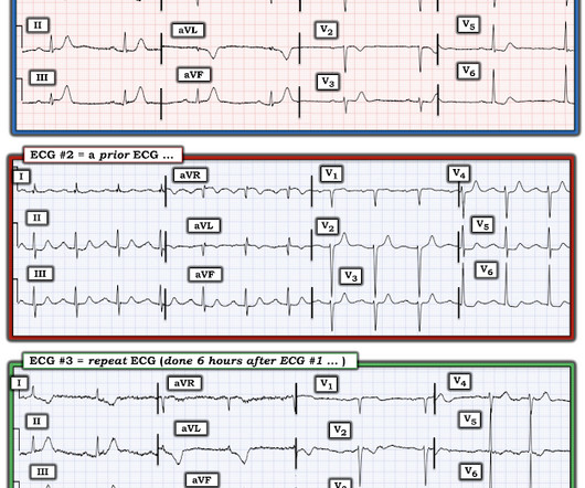
Written by Jesse McLaren A 70 year old with prior MIs and stents to LAD and RCA presented to the emergencydepartment with 2 weeks of increasing exertional chest pain radiating to the left arm, associated with nausea. 1] European guidelines add "regardless of biomarkers".
Upon arrival to the emergencydepartment, a senior emergency physician looked at the ECG and said "Nothing too exciting." Just because you don't see hemodynamically significant CAD on angiogram does not mean it is not OMI. V1 has 0.5 mm of elevation. More notably there are hyperacute T waves in V3 through V5.
Patient stated that he has had glucose over 400 even though he has not missed any doses of insulin. Aslanger's is a combination of inferior OMI with widespread ST depression and is due to BOTH occlusion of one artery (usually the circumflex, but sometimes the RCA) AND simultantous 3 vessel disease.
She was unable to be defibrillated but was cannulated and placed on ECMO in our EmergencyDepartment (ECLS - extracorporeal life support). Diagnosis of MINOCA should be made according to the Fourth Universal Definition of MI, in the absence of obstructive coronary artery disease (CAD) (no lesion ≥50%). The K was normal.
A 59-year-old male with a past medical history of a repaired ventricular septal defect (VSD), dextrocardia, hypertension, hyperlipidemia, and current smoker presented to the emergencydepartment (ED). 1 It has a prevalence of 0.01
At the bottom of the post, I have re-printed the section on aVR in my article on the ECG in ACS from the Canadian Journal of Cardiology: New Insights Into the Use of the 12-Lead Electrocardiogram for Diagnosing Acute Myocardial Infarction in the EmergencyDepartment Case 1. Total LM occlusion can present with STE or STD in aVR.
So I went to look at the chart and here is the history: This patient with no h/o CAD had a couple of episodes of chest pain during the day, then presented with one hour of substernal chest pain that had some reproducibility but also improved from 10/10 to 5/10 with nitroglycerine. Ann Emerg Med 1998;31(1):3-11.
Submitted and written by Alex Bracey with edits by Pendell Meyers and Steve Smith Case A 50ish year old man with a history of CAD w/ prior LAD MI s/p LAD stenting presented to the ED with chest pain similar to his prior MI, but worse. The pain initially started the day prior to presentation. The ST elevation from today is ~0.2
IIa C Pre-hospital logistics Management Recommendation Level of evidence The pre-hospital care of STEMI patients should be organized regionally (including all components from the emergency medical dispatch to catheterization laboratory) in order to provide reperfusion therapy as early as possible. I C Pain Titrated i.v.
Written by Pendell Meyers A man in his late 30s with history of hypertension, tobacco use, and obesity presented to the EmergencyDepartment for acute chest pain which started approximately 3 hours prior to arrival, in the setting of a very stressful situation. Scattered other nonobstructive CAD. You will get 5 free reports.
Written by Jesse McLaren, with comments from Smith An 85 year old with a history of CAD presented with 3 hours of chest pain that feels like heartburn but that radiates to the left arm. Below is the ECG. What do you think? There’s sinus bradycardia, first degree AV block, normal axis, delayed R wave progression, and normal voltages.
She presented to the EmergencyDepartment at around 3.5 Whether an ECG shows a pattern of OMI, SEI, both, or neither, the patient with ongoing ischemia needs to be considered for emergent reperfusion therapy. The chest pain was described as severe pressure radiating to both shoulders. Vital signs were within normal limits.
These are very commonly encountered in the emergencydepartment, so being able to correctly identify the rhythm is extremely important. He has a history of CHF, dilated cardiomyopathy, HTN, HLD and CAD. Lets dive in! When you are presented with a tachycardic ECG, we want you to focus on two major factors right away.
Case A 68 year old man with a medical history of hypertension, hyperlipidemia, and CAD with stent deployment in the RCA presented to the emergencydepartment with chest pain. He was worked up non-emergently in the ED with pain recurring and resolving multiple times during his stay. He had an EKG recorded right away.
We organize all of the trending information in your field so you don't have to. Join 5,000+ users and stay up to date on the latest articles your peers are reading.
You know about us, now we want to get to know you!
Let's personalize your content
Let's get even more personalized
We recognize your account from another site in our network, please click 'Send Email' below to continue with verifying your account and setting a password.


Let's personalize your content