This site uses cookies to improve your experience. To help us insure we adhere to various privacy regulations, please select your country/region of residence. If you do not select a country, we will assume you are from the United States. Select your Cookie Settings or view our Privacy Policy and Terms of Use.
Cookie Settings
Cookies and similar technologies are used on this website for proper function of the website, for tracking performance analytics and for marketing purposes. We and some of our third-party providers may use cookie data for various purposes. Please review the cookie settings below and choose your preference.
Used for the proper function of the website
Used for monitoring website traffic and interactions
Cookie Settings
Cookies and similar technologies are used on this website for proper function of the website, for tracking performance analytics and for marketing purposes. We and some of our third-party providers may use cookie data for various purposes. Please review the cookie settings below and choose your preference.
Strictly Necessary: Used for the proper function of the website
Performance/Analytics: Used for monitoring website traffic and interactions
A 45-year-old male with a history of chronic obstructive pulmonary disease (COPD), asthma, amphetamine and tetrahydrocannabinol (THC) use, and coronary vasospasm presented to triage with chest pain. During assessment, the patient reported that a left heart catheterization six months prior indicated spasms but no coronary artery disease.
They started CPR. After 1 mg of epinephrine they achieved ROSC. Total prehospital meds were epinephrine 1 mg x 3, amiodarone 300 mg and 100 mL of 8.4% But cardiac arrest is a period of near zero flow in the coronary arteries and causes SEVERE ischemia. It also does not uniformly indicate severe coronary disease.
Emergent coronary angiography is not recommended over a delayed or selective strategy in patients with ROSC after cardiac arrest in the absence of ST-segment elevation, shock, electrical instability, signs of significant myocardial damage, and ongoing ischemia (Level 3: no benefit). COR 1, LOE B-R. COR 2a, LOE B-R. COR 2a, LOE C-LD.
There was no bystander CPR. link] __ Case continued There was hypotension, initially controlled with an epinephrine infusion. For this reason we did not believe this was an acute coronary event and did not activate the cath lab. So a dual chamber pacer is placed with one lead through the coronary sinus to the LV.
It was witnessed, and CPR was performed by trained individuals. Fine ventricular fibrillation She received 2 mg epinephrine, 150 mg amiodarone and underwent chest compressions with the LUCAS device. Fine ventricular fibrillation She received 2 mg epinephrine, 150 mg amiodarone and underwent chest compressions with the LUCAS device.
CPR was started immediately. She was given 3 mg IV epinephrine and multiple rounds of ACLS over approximately 20 minutes. This is commonly found after epinephrine for cardiac arrest, but could have been pre-existing and a possible contributing factor to cardiac arrest. EMS arrived and found her in a wide complex PEA rhythm.
He underwent CPR, and regained a pulse after epinephrine, with an organized narrow complex rhythm at 140, but still with severe shock. And so it is wise to look at the coronary arteries. This ECG certainly looks like myocarditis, and was due to myocarditis, but missing acute coronary occlusion is not acceptable.
Medics found her apneic and pulseless, began CPR, and she was found to be in asystole. With ventilations and epinephrine, she regained a pulse. Rather it is due to coronary insufficiency due to a tight left main or 3-vessel disease with inadequate coronary flow.
CPR was initiated immediately. As in all ischemia interpretations with OMI findings, the findings can be due to type 1 AMI (example: acute coronary plaque rupture and thrombosis) or type 2 AMI (with or without fixed CAD, with severe regional supply/demand mismatch essentially equaling zero blood flow).
1 The primary goal of cardiopulmonary resuscitation (CPR) is to optimize coronary perfusion pressure and maintain systemic perfusion in order to prevent neurologic and other end-organ damage while working to achieve ROSC. There was no significant difference in cooling method between original cohorts.
We could not resuscitate her, but we did have excellent perfusion with LUCAS CPR, such that pulse oximetry had excellent waveform and 100% saturations, end tidal CO2 was 35, and cerebral perfusion monitoring was near normal throughout the attempted resuscitation. Armstrong, MD Arch Intern Med. 1987;147(3):465-469. doi:10.1001/archinte.1987.00370030069014.
Reviewed by: Vicki Currie Article 5: Does occluding the femoral artery during neonatal CPR increase the likelihood of ROSC? (In The goal of chest compressions during neonatal resuscitation is to increase cerebral and coronary blood flow with the intention to achieve a return of spontaneous circulation (ROSC).
He was given 50 mcg epinephrine with good response in both heart rate and blood pressure. His heart rate had improved to the 80s after epinephrine administration. CPR was initiated and he underwent 1 round of ACLS (CPR + 1 mg epi). During CPR, he started moving all four extremities spontaneously.
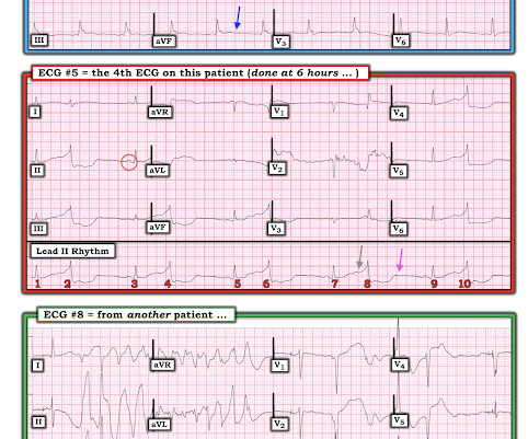
Angiography was performed at 10:31, just under 13 hours after the patients ED presentation: The red arrow shows a 50% distal stenosis of the left main coronary artery involving the ostium of the LAD. He suffered another cardiac arrest in the ICU with ROSC after another dose of epinephrine and one round of CPR.
We organize all of the trending information in your field so you don't have to. Join 5,000+ users and stay up to date on the latest articles your peers are reading.
You know about us, now we want to get to know you!
Let's personalize your content
Let's get even more personalized
We recognize your account from another site in our network, please click 'Send Email' below to continue with verifying your account and setting a password.


Let's personalize your content