This site uses cookies to improve your experience. To help us insure we adhere to various privacy regulations, please select your country/region of residence. If you do not select a country, we will assume you are from the United States. Select your Cookie Settings or view our Privacy Policy and Terms of Use.
Cookie Settings
Cookies and similar technologies are used on this website for proper function of the website, for tracking performance analytics and for marketing purposes. We and some of our third-party providers may use cookie data for various purposes. Please review the cookie settings below and choose your preference.
Used for the proper function of the website
Used for monitoring website traffic and interactions
Cookie Settings
Cookies and similar technologies are used on this website for proper function of the website, for tracking performance analytics and for marketing purposes. We and some of our third-party providers may use cookie data for various purposes. Please review the cookie settings below and choose your preference.
Strictly Necessary: Used for the proper function of the website
Performance/Analytics: Used for monitoring website traffic and interactions
After 1 mg of epinephrine they achieved ROSC. Total prehospital meds were epinephrine 1 mg x 3, amiodarone 300 mg and 100 mL of 8.4% But cardiac arrest is a period of near zero flow in the coronary arteries and causes SEVERE ischemia. It also does not uniformly indicate severe coronary disease. sodium bicarbonate.
2 Standard management for VT and VF involves the use of electrical defibrillation, high-quality chest compressions, and epinephrine. 5 More recent literature defines “refractory” as VT or VF that is persistent or recurrent despite three shocks from a defibrillator, three rounds of epinephrine, and use of an antiarrhythmic (i.e.,
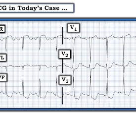
In the ED he received methylprednisolone, diphenhydramine, and epinephrine for possible anaphylaxis. Shortly after receiving epinephrine, the patient developed new leg cramps and chest pain. Meyers — No definitive explanation for the marked ST segment deviation seen in ECG #1 was determined.
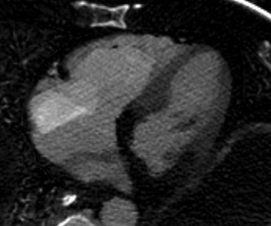
It shows a proximal LAD occlusion, in conjunction with a subtotally occluded LMCA ( Left Main Coronary Artery ). Epinephrine infusion was begun. Upon contrast injection of the LMCA, the patient deteriorated, as the LMCA was severely diseased and flow to all coronary arteries ( LAD, LCx and RCA ) was compromised.
Fine ventricular fibrillation She received 2 mg epinephrine, 150 mg amiodarone and underwent chest compressions with the LUCAS device. The patient was taken for an angiogram and had an 80% LAD lesion, but it could not be definitely determined whether this was an acute thrombotic lesion or a chronic stable lesion. It was stented.
With ventilations and epinephrine, she regained a pulse. Rather it is due to coronary insufficiency due to a tight left main or 3-vessel disease with inadequate coronary flow. A middle-age woman with h/o hypertension was found down by her husband. She was never seen to be in ventricular fibrillation and was never defibrillated.
She was given 3 mg IV epinephrine and multiple rounds of ACLS over approximately 20 minutes. This is commonly found after epinephrine for cardiac arrest, but could have been pre-existing and a possible contributing factor to cardiac arrest. The ultimate reason for the long QT was never definitively determined.
They stated that the patient was coded for 20 minutes, including multiple doses of epinephrine, and they also gave glucose, calcium, and bicarb. As stated above, resuscitation included epinephrine, calcium, and bicarb. Of course this must be followed immediately with definitive therapies and potassium source control if possible.
The diagnostic coronary angiogram identified only minimal coronary artery disease, but there was a severely calcified, ‘immobile’ aortic valve. Author continued : STE in aVR is often due to left main coronary artery obstruction (OR 4.72), and is associated with in-hospital cardiovascular mortality (OR 5.58).
Limitations Single center study in India A very specific subset of patients with advanced liver cirrhosis and non-variceal upper GI bleeding (gastritis, portal hypertensive gastropathy, ulcers) Investigators also excluded patients on antiplatelet and anticoagulant therapy, likely eliminating many patients with diabetes and coronary artery disease.
He was given 50 mcg epinephrine with good response in both heart rate and blood pressure. His heart rate had improved to the 80s after epinephrine administration. He was persistently bradycardic, requiring 2 x 50 mcg epinephrine to maintain HR >60. Epinephrine drip was started and norepinephrine was discontinued.
We organize all of the trending information in your field so you don't have to. Join 5,000+ users and stay up to date on the latest articles your peers are reading.
You know about us, now we want to get to know you!
Let's personalize your content
Let's get even more personalized
We recognize your account from another site in our network, please click 'Send Email' below to continue with verifying your account and setting a password.


Let's personalize your content