This site uses cookies to improve your experience. To help us insure we adhere to various privacy regulations, please select your country/region of residence. If you do not select a country, we will assume you are from the United States. Select your Cookie Settings or view our Privacy Policy and Terms of Use.
Cookie Settings
Cookies and similar technologies are used on this website for proper function of the website, for tracking performance analytics and for marketing purposes. We and some of our third-party providers may use cookie data for various purposes. Please review the cookie settings below and choose your preference.
Used for the proper function of the website
Used for monitoring website traffic and interactions
Cookie Settings
Cookies and similar technologies are used on this website for proper function of the website, for tracking performance analytics and for marketing purposes. We and some of our third-party providers may use cookie data for various purposes. Please review the cookie settings below and choose your preference.
Strictly Necessary: Used for the proper function of the website
Performance/Analytics: Used for monitoring website traffic and interactions
On ED arrival GCS is 3, there are rapid eye movements to the right but no other apparent seizure activity. Propofol utilized for sedation; patient admitted to ICU for EEG monitoring. Official diagnosis requires EEG, which is not something we can typically obtain in the ED. They administer two doses of 10 mg midazolam IM.
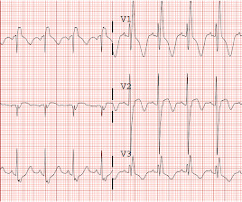
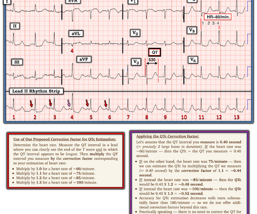
A 65 y/o Female was admitted to the ICU for septic shock. The combination of prolonged QT and deep T wave inversion throughout the precordium is typical of Takotsubo syndrome, or Stress Cardiomyopathy – which can occur in the context of a physiologically distressed ICU patient, further compromising their hemodynamics. Surawicz, B.
This is critical for the EMS provider, or ED clinician, as identification of Grade I ischemia (aka, HATW’s) addresses the culprit lesion at the earliest opportunity with excellent downstream prognosis for the patient. [2] The following ECG was captured upon arrival at the receiving ED. The ED resulted an 8.7 An ECG is recorded.
The patient was upgraded to the ICU for closer monitoring. Electrocardiographic Differentiation Between Acute Pulmonary Embolism and Acute Coronary Syndromes on the Basis of Negative T Waves - ScienceDirect. Echocardiogram showed severe RV dilation with McConnell’s sign and an elevated RVSP. In fact, Kosuge et al.
The pacing rate was increased without clinical improvement and the patient was transferred to the ICU for closer monitoring/treatment. The patient is an older woman with known coronary disease and an ICD-Pacemaker implanted because of a history of VT ( V entricular T achycardia ). small squares in width (260ms).
A temporary pacemaker was implanted, and she was admitted to the ICU with cardiogenic shock. I interpreted this tracing knowing only that the patient was a woman in her 60s, with a prior history of proximal LAD OMI — who now presented to the ED with a history of new chest discomfort and shortness of breath.
A 70-year-old female with a past medical history of hypertension, coronary artery disease s/p 2x drug eluting stent placement one month ago, atrial fibrillation on apixaban presents to the ED with weakness and lightheadedness. A 25-year-old man presents to the ED via EMS after he sustained a gunshot wound to the left flank.
Course : A CT of the head, neck, chest, abdomen and pelvis showed no other unanticipated injuries and she was admitted to the ICU. More commonly, however, ECG and/or echo wall motion abnormalities are due to pre-existing disease, especially coronary disease complicating trauma.
The patient was admitted to the ICU for close monitoring and electrolyte repletion and had an uneventful hospital course. The patient is a man in his 60s with established severe alcohol use disorder — and epidural abscess being treated with longterm Ciprofloxacin — who presented to the ED following a syncopal episode. As per Drs.
mg/dL (sorry, Europeans, for the weird units) Here was the initial ED ECG: There is a junctional rhythm with retrograde P-waves (see the dip in the T-wave in lead II across the bottom; you can follow that up to all the other leads and see the retrograde P wave). He was admitted to the ICU and was unstable, in shock, overnight.
A 68-year-old male with a past medical history of hypertension, diabetes mellitus, and coronary artery disease with a drug eluting stent placed 2 months ago presents with dizziness and vomiting that began 3 hours ago. Median time from ED arrival to diagnosis was 8 hours 24 min in one study, with only 19% being diagnosed within the 4.5-hour
The patient vomited once and given the more intense pain decided to come to the ED. Immediate and early percutaneous coronary intervention in very high-risk and high-risk Non-STEMI patients. 2-hour hsTn: 615 ng/L; bedside ED echo (without contrast) did not show a clear wall motion abnormality (WMA). Lupu L, et al. mg/dL, K 3.5
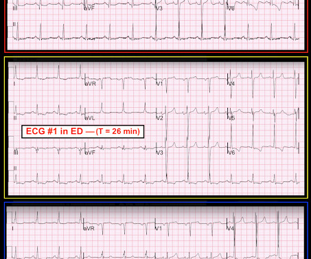
This is her pre-hospital ECG: This is her first ECG in the ED: What do you think? She also received an additional nitro in the ED after receiving aspirin and nitro via EMS. If she had no risk factors, it is doubtful that she would have developed such extensive coronary artery disease as we see on the angiogram.
in the ICU but survived with excellent function. IF , on the other hand — the patient with new chest pain is older and lacks predisposing viral symptoms — then acute pericarditis becomes a rare diagnosis in an ED setting. I completely agree with many of the KEY findings conveyed by Dr. Meyers.
Upon arrival in the ICU, before getting Continuous Veno-Venous Hemodialysis (CVVHD), his potassium had risen again to 7.8 Acute coronary syndrome is unlikely to be one of those entities. Bottom Line: Hyperkalemia is an increasingly common ED diagnosis that must not be missed. There is no ECG available from this time.
She was asymptomatic at the time of this ECG recorded on arrival to our ED: What do you think? If for some reason the angiogram is delayed, they should receive maximal medical therapy in an ICU setting with continuous 12-lead ST segment monitoring under the close attention of a practitioner with advanced ECG interpretation training.
His ED cardiac ultrasound (which is not at all ideal for detecting wall motion abnormalities, and is also very operator dependent for this finding) was significant for depressed global EF. Fortunately, he was extubated several days later in the ICU with intact baseline mental status and was discharged shortly thereafter to subacute rehab.
We obtained access and monitoring, but she showed no signs of improvement, and we judged that an intervention must be done in the ED without delay. A CT was obtained later and showed appropriate positioning of the catheter: She was admitted to the ICU and the catheter was used several times to withdraw more fluid.
A 64-year-old male presents by EMS to the ED with shortness of breath. 1 There are over 50,000 visits related to heart transplant in the United States each year and over half of these patients are admitted to the hospital from the ED. We’ll keep it short, while you keep that EM brain sharp.
Some of the critical differentials include pulmonary embolism, acute decompensated heart failure, pneumonia, pneumothorax, and acute coronary syndrome. Anginal chest pain, chest heaviness, or evidence of fluid overload suggest acute coronary syndrome or acute decompensated heart failure. Signs and symptoms of systemic infection (e.g.,
Fifth , potential management actions are in your hands; you do not need to request a coronary interventionalist or cath lab team. Making a specific ECG Diagnosis (less important in the ED) Without reading the below, I suspected posterior fascicular VT. Again the clinical history is helpful.
A 43-year-old male with a history of mitral valve regurgitation s/p valvular replacement, hypertension, hyperlipidemia was evaluated in the ED for septic shock secondary to a pyelonephritis with a renal abscess. Left ventricular outflow tract obstruction in ICU patients. He has clinically deteriorated and required intubation.
This is likely because Dexmed helps dampen the sympathetic response to perioperative stress, improving coronary artery perfusion. How is Dexmedetomidine used in the ED? Future Roles of Dexmedetomidine in the Emergency Department Dexmedetomidine is increasingly being explored as a paediatric procedural sedation option in the ED.
We organize all of the trending information in your field so you don't have to. Join 5,000+ users and stay up to date on the latest articles your peers are reading.
You know about us, now we want to get to know you!
Let's personalize your content
Let's get even more personalized
We recognize your account from another site in our network, please click 'Send Email' below to continue with verifying your account and setting a password.


Let's personalize your content