This site uses cookies to improve your experience. To help us insure we adhere to various privacy regulations, please select your country/region of residence. If you do not select a country, we will assume you are from the United States. Select your Cookie Settings or view our Privacy Policy and Terms of Use.
Cookie Settings
Cookies and similar technologies are used on this website for proper function of the website, for tracking performance analytics and for marketing purposes. We and some of our third-party providers may use cookie data for various purposes. Please review the cookie settings below and choose your preference.
Used for the proper function of the website
Used for monitoring website traffic and interactions
Cookie Settings
Cookies and similar technologies are used on this website for proper function of the website, for tracking performance analytics and for marketing purposes. We and some of our third-party providers may use cookie data for various purposes. Please review the cookie settings below and choose your preference.
Strictly Necessary: Used for the proper function of the website
Performance/Analytics: Used for monitoring website traffic and interactions
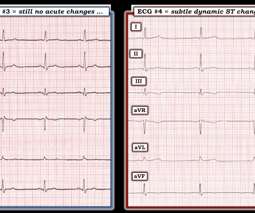
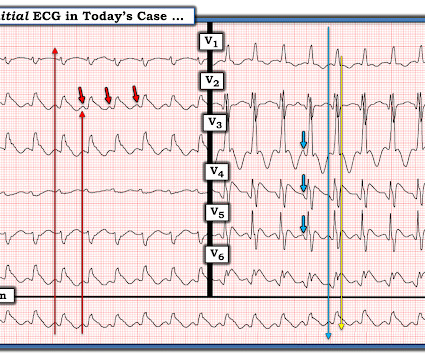
By Smith, peer-reviewed by Interventional Cardiologist Emre Aslanger Submitted by anonymous A 53 y.o. Here is his ED ECG at triage: Obvious high lateral OMI that does not quite meet STEMI criteria. male presents to the ED at 6:45 AM with left sided chest dull pressure that woke him up from sleep at 3am. He was started on nitro gtt.
He is interested and experienced in healthcare informatics, previously worked with ED-directed EMR design, and is involved in the New York City Health and Hospitals Healthcare Administration Scholars Program (HASP). Acute coronary syndrome (ACS) is responsible for the majority (60%) of all OHCAs in patients.
by Emre Aslanger Dr. Aslanger is our newest editorial member. Dr. Aslanger is also the author of the DIFFOCULT study: Emre K. Smith , d and Muzaffer Değertekin a DIFOCCULT: DIagnostic accuracy oF electrocardiogram for acute coronary OCClUsion resuLTing in myocardial infarction. He is an interventional cardiologist in Turkey.
Their OMI Manifesto details how use of standard STEMI criteria results in an unacceptable level of inaccuracy, in which an estimated 25-30% of acute coronary occlusions are missed! The article by Aslanger, Smith et al that is featured above in today’s post has just been published.
This was contributed by Co-editor Emre Aslanger, an interventional cardiologist in Turkey. A recent angiogram report indicated a totally occluded left anterior descending artery (LAD) and right coronary artery (RCA), with 30-40% narrowings in the left circumflex artery (LCx). That was also my initial concern.
I published, and Emre Aslanger externally validated, the 4-Variable formula for differentiating the ST Elevation of LAD OMI from Normal ST Elevation. Knowing the patient has a history of coronary disease could be relevant to today's case — as it should add to our suspicion of a new acute event.
Furthermore, some ECGs may not meet the STEMI criteria but may still be diagnostic for acute coronary occlusion (ACO). Smith and Emre Aslanger, but we also thank external researchers for their demonstrative ECGs (thanks to Philip L. The majority of the ECGs are from Stephen W. Mar for atrial activity ECG).
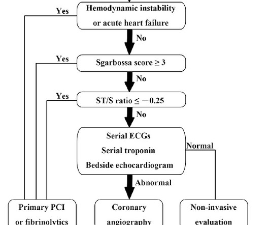
Now let’s compare this with the existing paradigm to identify multiple preventable delays to reperfusion, which can be improved through the paradigm shift from STEMI to OMI. In the STEMI paradigm, patients with ischemic symptoms and ECGs that don’t meet STEMI criteria get serial ECGs.
Hospital Course The patient was taken emergently to the cath lab which did not reveal any significant coronary artery disease, but she was noted to have reduced EF consistent with Takotsubo cardiomyopathy. Such cases are classified as MINOCA (Myocardial Infarction with Non-Obstructed Coronary Arteries). It can only be seen by IVUS.
The patient was brought to the ED as a possible Code STEMI and was seen directly by cardiology. Similarly, STEMI guidelines call for urgent angiography for refractory ischemia or electrical/hemodynamic instability, regardless of ECG findings. This ALONE is very strong evidence of acute coronary occlusion.
Written by Emre Aslanger. Emre is a new Editor of the Blog. Take home messages: Any coronary occlusion may present with vague symptoms, but when ECG is clear, there should not be any suspicion. PMID: 34523597. == MY Comment by K EN G RAUER, MD ( 11/13/2022 ): == Highly interesting case by Emre Aslanger. doi: 10.5543/tkda.2021.21026.
Written by Emre Aslanger (Emre is our newest editor. Although not striking, this is clearly a diagnostic ECG for infero"posterior" myocardial infarction due to coronary occlusion (OMI), most likely due to left circumflex (LCx) artery occlusion. Here are his publications.) He denies any illicit drug use. His ECG is shown below.
We organize all of the trending information in your field so you don't have to. Join 5,000+ users and stay up to date on the latest articles your peers are reading.
You know about us, now we want to get to know you!
Let's personalize your content
Let's get even more personalized
We recognize your account from another site in our network, please click 'Send Email' below to continue with verifying your account and setting a password.


Let's personalize your content