This site uses cookies to improve your experience. To help us insure we adhere to various privacy regulations, please select your country/region of residence. If you do not select a country, we will assume you are from the United States. Select your Cookie Settings or view our Privacy Policy and Terms of Use.
Cookie Settings
Cookies and similar technologies are used on this website for proper function of the website, for tracking performance analytics and for marketing purposes. We and some of our third-party providers may use cookie data for various purposes. Please review the cookie settings below and choose your preference.
Used for the proper function of the website
Used for monitoring website traffic and interactions
Cookie Settings
Cookies and similar technologies are used on this website for proper function of the website, for tracking performance analytics and for marketing purposes. We and some of our third-party providers may use cookie data for various purposes. Please review the cookie settings below and choose your preference.
Strictly Necessary: Used for the proper function of the website
Performance/Analytics: Used for monitoring website traffic and interactions
EMS arrived and found him in Ventricular Fibrillation (VF). After 1 mg of epinephrine they achieved ROSC. Total prehospital meds were epinephrine 1 mg x 3, amiodarone 300 mg and 100 mL of 8.4% But cardiac arrest is a period of near zero flow in the coronary arteries and causes SEVERE ischemia. They started CPR.
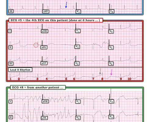
link] __ Case continued There was hypotension, initially controlled with an epinephrine infusion. For this reason we did not believe this was an acute coronary event and did not activate the cath lab. Here is the troponin profile overnight: This is consistent with cardiac arrest without acute coronary occlusion.
2 Standard management for VT and VF involves the use of electrical defibrillation, high-quality chest compressions, and epinephrine. 5 More recent literature defines “refractory” as VT or VF that is persistent or recurrent despite three shocks from a defibrillator, three rounds of epinephrine, and use of an antiarrhythmic (i.e.,
The patient in today’s case is a previously healthy 40-something male who contacted EMS due to acute onset crushing chest pain. It shows a proximal LAD occlusion, in conjunction with a subtotally occluded LMCA ( Left Main Coronary Artery ). Epinephrine infusion was begun. The below ECG was recorded.
They stated that the patient was coded for 20 minutes, including multiple doses of epinephrine, and they also gave glucose, calcium, and bicarb. As stated above, resuscitation included epinephrine, calcium, and bicarb. He had been given 3 grams Ca gluconate by EMS. Acute coronary syndrome is unlikely to be one of those entities.
It was edited by Smith CASE : A 52-year-old male with a past medical history of hypertension and COPD summoned EMS with complaints of chest pain, weakness and nausea. En route, EMS administered aspirin 325mg by mouth, but withheld nitroglycerin due to initial hypotension. Answer below in the still shot.
EMS arrived and found her in a wide complex PEA rhythm. She was given 3 mg IV epinephrine and multiple rounds of ACLS over approximately 20 minutes. This is commonly found after epinephrine for cardiac arrest, but could have been pre-existing and a possible contributing factor to cardiac arrest. CPR was started immediately.
As in all ischemia interpretations with OMI findings, the findings can be due to type 1 AMI (example: acute coronary plaque rupture and thrombosis) or type 2 AMI (with or without fixed CAD, with severe regional supply/demand mismatch essentially equaling zero blood flow). On epinephrine and norepinephrine drips."
Limitations Single center study in India A very specific subset of patients with advanced liver cirrhosis and non-variceal upper GI bleeding (gastritis, portal hypertensive gastropathy, ulcers) Investigators also excluded patients on antiplatelet and anticoagulant therapy, likely eliminating many patients with diabetes and coronary artery disease.
We’ll keep it short, while you keep that EM brain sharp. The catheterization lab is activated, but catheterization shows no coronary artery occlusion. A 67-year-old female with past medical history of hypertension presents with acute onset of chest pain without associated symptoms. Initial troponin is mildly elevated.
Resuscitated with chest compressions, epinephrine. including epinephrine, and there was ROSC. Moreover, it does not follow a coronary distribution very well. The coronaries were clean. PEA is uncommon as an initial rhythm witnessed by EMS on the scene when the cause is an acute ischemic event. From this site.
Authors: Rachel Bridwell, MD (EM Attending Physician; Tacoma, WA), Katey DG Osborne, MD (EM Attending Physician; Tacoma, WA) // Reviewed by: Alex Koyfman, MD (@EMHighAK, EM Attending Physician, UTSW / Parkland Memorial Hospital) and Brit Long, MD (@long_brit, EM Attending Physician, San Antonio, TX) Welcome to emDOCs revamp!
In the EMS setting, the most common cardiogenic shock patient is most likely a STEMI. The ultimate goal is to optimize coronary perfusion pressure (CPP)—in other words, the amount of blood flow into the coronary arteries. When the heart is full, it puts pressure on the myocardium, compressing the coronary microvasculature.
We organize all of the trending information in your field so you don't have to. Join 5,000+ users and stay up to date on the latest articles your peers are reading.
You know about us, now we want to get to know you!
Let's personalize your content
Let's get even more personalized
We recognize your account from another site in our network, please click 'Send Email' below to continue with verifying your account and setting a password.


Let's personalize your content