This site uses cookies to improve your experience. To help us insure we adhere to various privacy regulations, please select your country/region of residence. If you do not select a country, we will assume you are from the United States. Select your Cookie Settings or view our Privacy Policy and Terms of Use.
Cookie Settings
Cookies and similar technologies are used on this website for proper function of the website, for tracking performance analytics and for marketing purposes. We and some of our third-party providers may use cookie data for various purposes. Please review the cookie settings below and choose your preference.
Used for the proper function of the website
Used for monitoring website traffic and interactions
Cookie Settings
Cookies and similar technologies are used on this website for proper function of the website, for tracking performance analytics and for marketing purposes. We and some of our third-party providers may use cookie data for various purposes. Please review the cookie settings below and choose your preference.
Strictly Necessary: Used for the proper function of the website
Performance/Analytics: Used for monitoring website traffic and interactions
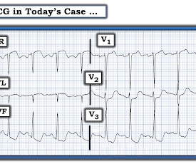
A 45-year-old male with a history of chronic obstructive pulmonary disease (COPD), asthma, amphetamine and tetrahydrocannabinol (THC) use, and coronary vasospasm presented to triage with chest pain. During assessment, the patient reported that a left heart catheterization six months prior indicated spasms but no coronary artery disease.
After 1 mg of epinephrine they achieved ROSC. Total prehospital meds were epinephrine 1 mg x 3, amiodarone 300 mg and 100 mL of 8.4% But cardiac arrest is a period of near zero flow in the coronary arteries and causes SEVERE ischemia. It also does not uniformly indicate severe coronary disease. sodium bicarbonate.
Emergent coronary angiography is not recommended over a delayed or selective strategy in patients with ROSC after cardiac arrest in the absence of ST-segment elevation, shock, electrical instability, signs of significant myocardial damage, and ongoing ischemia (Level 3: no benefit). COR 1, LOE B-R. COR 2a, LOE B-R. COR 2a, LOE C-LD.
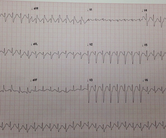
link] __ Case continued There was hypotension, initially controlled with an epinephrine infusion. For this reason we did not believe this was an acute coronary event and did not activate the cath lab. Here is the troponin profile overnight: This is consistent with cardiac arrest without acute coronary occlusion.
2 Standard management for VT and VF involves the use of electrical defibrillation, high-quality chest compressions, and epinephrine. 5 More recent literature defines “refractory” as VT or VF that is persistent or recurrent despite three shocks from a defibrillator, three rounds of epinephrine, and use of an antiarrhythmic (i.e.,
In the ED he received methylprednisolone, diphenhydramine, and epinephrine for possible anaphylaxis. Shortly after receiving epinephrine, the patient developed new leg cramps and chest pain. And , after resolution of the acute allergic reaction — underlying coronary disease was appropriately ruled out before discharge.
He was resuscitated with chest compressions and defibrillation and 1 mg of epinephrine. The next day, and angiogram showed normal coronary arteries. This young male had ventricular fibrillation during a triathlon. On his bib it stated that he had a congenital heart disorder. His initial ECG is shown here. He awoke and did well.
Potentiation of cocaine-induced coronary vasoconstriction by beta-adrenergic blockade Ann Int Med 897-903 112 12 1990 December 15 816 2 style='mso-element:field-end'> Indeed, major articles advising against the use of beta blockers in cocaine toxicity reference only this article and do not discuss cardioselectivity. Cigarroa, R.G. McBride, W.
Fine ventricular fibrillation She received 2 mg epinephrine, 150 mg amiodarone and underwent chest compressions with the LUCAS device. Updates on the Electrocardiogram in Acute Coronary Syndromes. Electrocardiogram patterns in acute left main coronary artery occlusion. The patient was discharged neurologically intact.
She was given 3 mg IV epinephrine and multiple rounds of ACLS over approximately 20 minutes. This is commonly found after epinephrine for cardiac arrest, but could have been pre-existing and a possible contributing factor to cardiac arrest. A 60-something woman presented after a witnessed cardiac arrest. CPR was started immediately.
The diagnostic coronary angiogram identified only minimal coronary artery disease, but there was a severely calcified, ‘immobile’ aortic valve. Author continued : STE in aVR is often due to left main coronary artery obstruction (OR 4.72), and is associated with in-hospital cardiovascular mortality (OR 5.58).
The ST segment changes are compatible with severe subendocardial ischemia which can be caused by type I MI from ACS or potentially from type II MI (non-obstructive coronary artery disease with supply/demand mismatch). This patient is actively dying from a left main coronary artery OMI and cardiac arrest from VT/VF or PEA is imminent!
It shows a proximal LAD occlusion, in conjunction with a subtotally occluded LMCA ( Left Main Coronary Artery ). Epinephrine infusion was begun. Upon contrast injection of the LMCA, the patient deteriorated, as the LMCA was severely diseased and flow to all coronary arteries ( LAD, LCx and RCA ) was compromised.
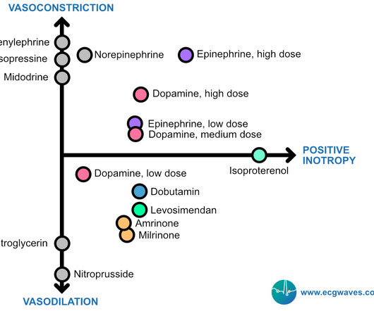
Below follows a drug manual for use in the CCU (coronary care unit), ICU (intensive care unit) or ER (emergency room). μg/kg/min + + + ++ Low dose dopamine stimulates D1 receptors and induces vasodilation in coronary, renal, cerebral and mesenteric vessels. Increases coronary blood flow. Coronary flow enhanced.
He underwent CPR, and regained a pulse after epinephrine, with an organized narrow complex rhythm at 140, but still with severe shock. And so it is wise to look at the coronary arteries. This ECG certainly looks like myocarditis, and was due to myocarditis, but missing acute coronary occlusion is not acceptable.
The patient has also developed sinus bradycardia, which may result from right coronary artery ischemia to the SA node. The patient is started on epinephrine infusion for cardiogenic shock and cardiology took the patient to the cath lab. The Queen of Hearts sees it of course: Still none of these three ECGs meet STEMI criteria.
They stated that the patient was coded for 20 minutes, including multiple doses of epinephrine, and they also gave glucose, calcium, and bicarb. As stated above, resuscitation included epinephrine, calcium, and bicarb. Acute coronary syndrome is unlikely to be one of those entities. After ROSC achieved: Sinus rhythm.
With ventilations and epinephrine, she regained a pulse. Rather it is due to coronary insufficiency due to a tight left main or 3-vessel disease with inadequate coronary flow. A middle-age woman with h/o hypertension was found down by her husband. She was never seen to be in ventricular fibrillation and was never defibrillated.
As in all ischemia interpretations with OMI findings, the findings can be due to type 1 AMI (example: acute coronary plaque rupture and thrombosis) or type 2 AMI (with or without fixed CAD, with severe regional supply/demand mismatch essentially equaling zero blood flow). On epinephrine and norepinephrine drips."
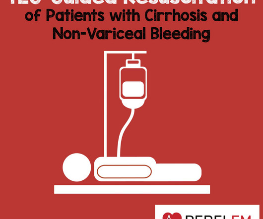
Limitations Single center study in India A very specific subset of patients with advanced liver cirrhosis and non-variceal upper GI bleeding (gastritis, portal hypertensive gastropathy, ulcers) Investigators also excluded patients on antiplatelet and anticoagulant therapy, likely eliminating many patients with diabetes and coronary artery disease.
This page summarises the most current recommendations for the management of acute coronary syndromes with persistent ST-segment elevations (i.e III A Primary percutaneous coronary intervention strategy Management Recommendation Level of evidence Primary PCI of the infarct related artery (IRA) is indicated.
Over a 13-month period, serum potassium and magnesium levels were measured in 590 patients admitted to a coronary care unit. Hypokalemia after cardiac arrest was independent of arterial pH, epinephrine or bicarbonate therapy during resuscitation, or prior therapy with diuretic drugs, digoxin or propranolol. 1987;147(3):465-469.
1 The primary goal of cardiopulmonary resuscitation (CPR) is to optimize coronary perfusion pressure and maintain systemic perfusion in order to prevent neurologic and other end-organ damage while working to achieve ROSC. Interventions during the acute phase of treatment post return of spontaneous circulation (ROSC) are therefore critical.
The catheterization lab is activated, but catheterization shows no coronary artery occlusion. ECG shows ST-segment elevation in V3-V6 only with depression in aVR. Initial troponin is mildly elevated. On further questioning, the patient denies recent illness but does mention that her daughter passed away in a car accident yesterday.
The goal of chest compressions during neonatal resuscitation is to increase cerebral and coronary blood flow with the intention to achieve a return of spontaneous circulation (ROSC). Both were partly delivered via caesarian section, flow probes inserted into carotid, pulmonary and left circumflex coronary arteries.
It eliminates the “pulse check,” allowing instant confirmation of pulsatility, while also allowing a very sophisticated assessment of coronary perfusion. * A diastolic BP above 3540 mmHg, measured from the arterial line during cardiac arrest, suggests adequate coronary perfusion.
He was given 50 mcg epinephrine with good response in both heart rate and blood pressure. His heart rate had improved to the 80s after epinephrine administration. He was persistently bradycardic, requiring 2 x 50 mcg epinephrine to maintain HR >60. Epinephrine drip was started and norepinephrine was discontinued.
The ultimate goal is to optimize coronary perfusion pressure (CPP)—in other words, the amount of blood flow into the coronary arteries. When the heart is full, it puts pressure on the myocardium, compressing the coronary microvasculature. When it gets too high, it can impair blood flow into the coronaries. mcg/kg/min.
EPINEPHRINE-INUDCED SHOCK: LEFT VENTRICULAR OUTFLOW TRACT OBSTRUCTION ON VASOPRESSORS. m/s)—problematic and elevated > 50 mm Hg (2.5 Left ventricular outflow tract gradient variability in hypertrophic cardiomyopathy. Clin Cardiol. 2009;32(7):397-402. doi:10.1002/CLC.20594 20594 MOZELL D, SHAH N, CASTILLON JJR, GONUGUNTLA VT, AWERBUCH E.
Resuscitated with chest compressions, epinephrine. including epinephrine, and there was ROSC. Moreover, it does not follow a coronary distribution very well. The coronaries were clean. Not a shockable rhythm. They laid her on the floor and called 911. Shortly thereafter, pulses were lost. This is unusual in acute OMI.
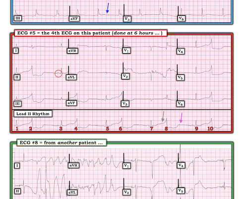
Angiography was performed at 10:31, just under 13 hours after the patients ED presentation: The red arrow shows a 50% distal stenosis of the left main coronary artery involving the ostium of the LAD. He suffered another cardiac arrest in the ICU with ROSC after another dose of epinephrine and one round of CPR. PCI was not performed.
We organize all of the trending information in your field so you don't have to. Join 5,000+ users and stay up to date on the latest articles your peers are reading.
You know about us, now we want to get to know you!
Let's personalize your content
Let's get even more personalized
We recognize your account from another site in our network, please click 'Send Email' below to continue with verifying your account and setting a password.


Let's personalize your content