This site uses cookies to improve your experience. To help us insure we adhere to various privacy regulations, please select your country/region of residence. If you do not select a country, we will assume you are from the United States. Select your Cookie Settings or view our Privacy Policy and Terms of Use.
Cookie Settings
Cookies and similar technologies are used on this website for proper function of the website, for tracking performance analytics and for marketing purposes. We and some of our third-party providers may use cookie data for various purposes. Please review the cookie settings below and choose your preference.
Used for the proper function of the website
Used for monitoring website traffic and interactions
Cookie Settings
Cookies and similar technologies are used on this website for proper function of the website, for tracking performance analytics and for marketing purposes. We and some of our third-party providers may use cookie data for various purposes. Please review the cookie settings below and choose your preference.
Strictly Necessary: Used for the proper function of the website
Performance/Analytics: Used for monitoring website traffic and interactions
His family started CPR and called EMS, who arrived to find him in ventricular fibrillation. Further information is not available. Despite anticipation by many that the initial post-resuscitation ECG will show an obvious acute infarction — this expected "STEMI picture" is often not seen.
The nurses started CPR immediately and place pads before you even arrived. A post-arrest ECG doesn’t show any signs of STEMI. For more information on the fragility index (FI) click on this LINK. The patient is in ventricular fibrillation, and you achieve return of spontaneous circulation (ROSC) on the second shock.
He reports that this chest pain feels different than prior chest pain when he had his STEMI/OMI, but is unable to further describe chest pain. I sent it to 5 of my OMI friends without any clinical information or outcome and all 5 independently responded with exactly the same diagnosis: "reperfused inferior OMI".
Did they get bystander CPR? We don’t know any of this information unfortunately and all are key in patient selection The median lactate level before revascularization was 6.9mmol/L (Range 4.6 Control: 53.4% D ECLS: 18.2% Control 8.7% Control 38.0% Control: 49.0% RR 0.98; 95% CI 0.80 to 1.19; p = 0.81 vs 13.9% (RR 0.58; 95% CI 0.33
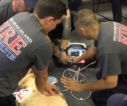
This is an extremely important topic especially for EMS systems that are implementing High Performance CPR , because it is very predictable that you are going to see a lot more patients with return of pulses in the field, and if you don’t have a plan, lots of things can go wrong before arriving at the hospital.
I sent this ECG with no clinical information to Dr. Smith. This rhythm reportedly produced no palpable pulse, and CPR was continued. 30 seconds later, however, the patient began spontaneously moving and CPR was discontinued. A repeat ECG was done: Obvious anterolateral wall STEMI.
Jesse McLaren (@ECGcases), of Emergency Medicine Cases Reviewed by Pendell Meyers and Steve Smith An 85yo with a history of hypertension developed chest pain and collapsed, and had bystander CPR. The patient was brought to the ED as a possible Code STEMI and was seen directly by cardiology. Learning points 1.
Might be a good idea to revisit this) You likely practice CPR, cannulation, airway insertion and all the fun stuff, but I guarantee that you will spend more time talking on the radio than doing those things. Not only that, but the information they want to give you may be important. Learn it, use it.
Here is his triage ECG: PM Cardio version: With no other information at all, I sent this ECG to Dr. Smith, who replied: "I think it is real. He underwent CPR and then was shocked out of VF. Like Dr. Smith — I was shown this patient's initial ECG without the benefit of any clinical information. STD in V4-5 too."
Evaluate and treat seizures or SE after CA in the context of other available clinical information because other systemic factors may influence the occurrence of seizures or SE and the effectiveness of treatment (90%, 18/20). The treatment goal for post-CA SE is seizure suppression or burst suppression for a minimum of 24 hours (95%, 19/20).
CPR was initiated immediately. I sent it to 2 of my ECG nerd colleagues with no clinical information whatsoever, who instantly said: "Looks like afib with subendocardial ischemia and right heart strain pattern." "I It was reportedly a PEA arrest; there was no recorded V Fib and no defibrillation.
He underwent CPR, and regained a pulse after epinephrine, with an organized narrow complex rhythm at 140, but still with severe shock. In a series of 18 patients with COVID and ST elevation, 8 were diagnosed with STEMI, 6 of whom had an angiogram and it showed obstructive coronary disease. He was intubated and then went pulseless.
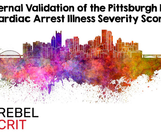
Background Information: Multiple illness severity scores have been developed for use after out-of-hospital cardiac arrest (OHCA) and in-hospital cardiac arrest (IHCA). Unfortunately, these rely on information that is not immediately available to providers in the early hours following return of spontaneous circulation (ROSC).
Angiography was technically challenging as the patient was receiving CPR, but the cardiologist suspected acute stent thrombosis and initiated cangrelor, although no repeat angiography was able to be obtained. There was indication of parasympathetic overdrive ( the acute inferior STEMI with profound bradycardia and junctional escape ).
We organize all of the trending information in your field so you don't have to. Join 5,000+ users and stay up to date on the latest articles your peers are reading.
You know about us, now we want to get to know you!
Let's personalize your content
Let's get even more personalized
We recognize your account from another site in our network, please click 'Send Email' below to continue with verifying your account and setting a password.


Let's personalize your content