This site uses cookies to improve your experience. To help us insure we adhere to various privacy regulations, please select your country/region of residence. If you do not select a country, we will assume you are from the United States. Select your Cookie Settings or view our Privacy Policy and Terms of Use.
Cookie Settings
Cookies and similar technologies are used on this website for proper function of the website, for tracking performance analytics and for marketing purposes. We and some of our third-party providers may use cookie data for various purposes. Please review the cookie settings below and choose your preference.
Used for the proper function of the website
Used for monitoring website traffic and interactions
Cookie Settings
Cookies and similar technologies are used on this website for proper function of the website, for tracking performance analytics and for marketing purposes. We and some of our third-party providers may use cookie data for various purposes. Please review the cookie settings below and choose your preference.
Strictly Necessary: Used for the proper function of the website
Performance/Analytics: Used for monitoring website traffic and interactions
After administering 1mg of epinephrine ROSC is noted with a bradycardic rhythm ( Figure 2 ). On ED arrival ROSC is achieved. As this case shows, electrical capture isn't always possible at lower currents, especially with pads placed in a standard anterolateral "defibrillation" position.
He was defibrillated into VT. He then underwent dual sequential defibrillation into asystole. After 1 mg of epinephrine they achieved ROSC. Total prehospital meds were epinephrine 1 mg x 3, amiodarone 300 mg and 100 mL of 8.4% The patient was brought to the ED and had this ECG recorded: What do you think?
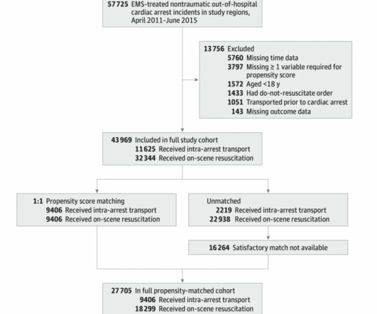
emergency departments (EDs), with statistics reporting more than 356,000 out-of-hospital cardiac arrests per year. 2 Standard management for VT and VF involves the use of electrical defibrillation, high-quality chest compressions, and epinephrine. Out-of-hospital cardiac arrest is a commonly encountered entity in U.S.
He was defibrillated, but they also noticed that he was being internally defibrillated and then found that he had an implantable ICD. He was unidentified and there were no records available After 7 shocks, he was successfully defibrillated and brought to the ED. Here is the initial ED ECG. What do you think?
This post will focus on the key parts of the guideline that affect ED evaluation and management. Vasopressor medications during cardiac arrest We recommend that epinephrine be administered for patients in cardiac arrest. It is reasonable to administer epinephrine 1 mg every 3 to 5 minutes for cardiac arrest. COR 1, LOE B-R.
Case: A 6-month-old boy presents to the emergency department (ED) with three days of worsening cough, cold symptoms, and fever. Background: We often manage patients in cardiac arrest in the ED or the intensive care unit (ICU). Pediatric Crit Care Med. Parents note that he has been progressively more tired and difficult to arouse.
Case: During a busy emergency department (ED) shift the paramedic phone rings. This has included things like therapeutic hypothermia ( SGEM#54 , SGEM#82 , SGEM#183 and SGEM#275 ), supraglottic devices ( SGEM#247 ), crowd sourcing CPR ( SGEM#143 and SGEM#306 ), and epinephrine ( SGEM#238 ).
Louis) // Reviewed by: Alex Koyfman, MD (@EMHighAK); Brit Long, MD (@long_brit) Case You are working in the trauma/critical care pod of your emergency department (ED). His blood sugar was normal en route to the ED, and his initial rhythm on the cardiac monitor was asystole. It is unclear how long he was down.
After the fourth defibrillation attempt, 200 mcg IV NTG was administered, resulting in immediate return of spontaneous circulation with a junctional bradycardia rhythm. Traditional Advanced Cardiovascular Life Support (ACLS) medications, namely epinephrine, have been known to exacerbate coronary vasospasm. Click to enlarge.)
The arrhythmia spontaneously converted before defibrillation was achieved. The patient was administered thrombolytics and shortly after the lytics were administered, the systolic blood pressure rose to about 80mmHg with ongoing epinephrine infusion. Those who make it to the ED usually have transient occlusions with reperfusion.
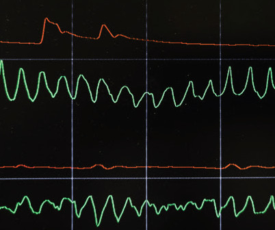
She was found to be in ventricular fibrillation and was defibrillated 8 times without a single, even transient, conversion out of fibrillation. Fine ventricular fibrillation She received 2 mg epinephrine, 150 mg amiodarone and underwent chest compressions with the LUCAS device. at the time of the ECG. References : 1.
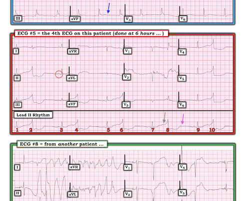
She was given 3 mg IV epinephrine and multiple rounds of ACLS over approximately 20 minutes. She was never defibrillated. I was texted this ECG in real time, but it turns out to actually be the 2nd one recorded in the ED. ECG #4 — This is the 3rd ECG that was done in this case ( obtained ~1 hour after arrival in the ED ).
With ventilations and epinephrine, she regained a pulse. She was never seen to be in ventricular fibrillation and was never defibrillated. She was hypotensive in the ED and her bedside echo showed a normal RV and LV. Data collected included demographics, initial rhythm, EKG, emergency department (ED) CT and outcomes.
He reportedly told his family "I think I'm having a heart attack", then they immediately drove him to the ED, and he was able to ambulate into the triage area before he collapsed and became unresponsive. It was reportedly a PEA arrest; there was no recorded V Fib and no defibrillation. On epinephrine and norepinephrine drips."
Here is his ED ECG: There is obvious infero-posterior STEMI. At cath, he immediately had incessant Torsades de Pointes requiring defibrillation 7 times and requiring placement of a transvenous pacer for overdrive pacing at a rate of 80. Medics stated that he had not been taking his clopidogrel for 2 weeks. He appeared to be in shock.
A 67-year-old man presents to the emergency department (ED) in cardiac arrest. Multiple attempts at defibrillation, epinephrine, and amiodarone have been unsuccessful. On ED presentation, he is unresponsive and the monitor shows ventricular fibrillation. He was found by bystanders after he collapsed and 911 was called.
EMS report was that the patient had unknown down time with unwitnessed arrest, found initially in VFib arrest, defibrillated x1 followed by PEA arrest alternating with asystolic arrest during transport. Chest compressions were continued, and the patient was given 1 round of epinephrine, calcium, bicarb, glucose.
Holy Foley A Rare Case of Iatrogenic Obstruction by Adam Heilmann, MD; Jessica Pelletier, DO; Jennifer Reyes Lin, MD, MPH Our patient is a 33-year-old male with spastic quadriparesis due to cerebral palsy with chronic indwelling suprapubic catheter (SPC) who presented to the emergency department (ED) due to concern for Foley catheter obstruction.
We talk about the nitty-gritty details of a well-run cardiac arrest, with Scott Weingart of Emcrit (@emcrit), ED intensivist. Takeaway lessons Resources We talk about the nitty-gritty details of a well-run cardiac arrest, with Scott Weingart of Emcrit ( @emcrit ), ED intensivist. Learn more at the Intensive Care Academy!
This is what the providers in the ED understood on patient arrival: Patient called 911 for syncope, then had witnessed PEA arrest after medics arrived. Resuscitated with chest compressions, epinephrine. including epinephrine, and there was ROSC. Not a shockable rhythm. Resuscitation was begun with chest compressions, etc.,
We organize all of the trending information in your field so you don't have to. Join 5,000+ users and stay up to date on the latest articles your peers are reading.
You know about us, now we want to get to know you!
Let's personalize your content
Let's get even more personalized
We recognize your account from another site in our network, please click 'Send Email' below to continue with verifying your account and setting a password.


Let's personalize your content