This site uses cookies to improve your experience. To help us insure we adhere to various privacy regulations, please select your country/region of residence. If you do not select a country, we will assume you are from the United States. Select your Cookie Settings or view our Privacy Policy and Terms of Use.
Cookie Settings
Cookies and similar technologies are used on this website for proper function of the website, for tracking performance analytics and for marketing purposes. We and some of our third-party providers may use cookie data for various purposes. Please review the cookie settings below and choose your preference.
Used for the proper function of the website
Used for monitoring website traffic and interactions
Cookie Settings
Cookies and similar technologies are used on this website for proper function of the website, for tracking performance analytics and for marketing purposes. We and some of our third-party providers may use cookie data for various purposes. Please review the cookie settings below and choose your preference.
Strictly Necessary: Used for the proper function of the website
Performance/Analytics: Used for monitoring website traffic and interactions
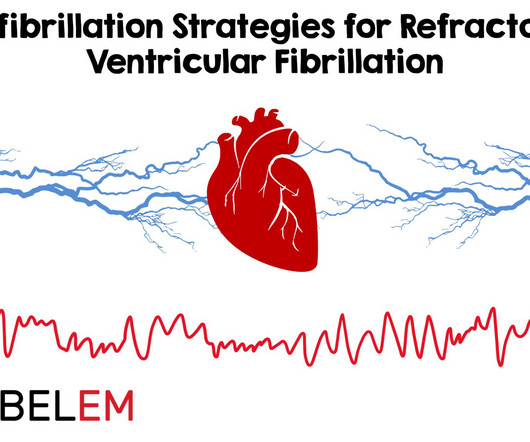
Background Information: Double external defibrillation (DED) is an intervention often used to treat refractory ventricular fibrillation (RVF). This procedure involves applying another set of pads attached to a second defibrillator to a patient and shocking them in hopes of terminating the rhythm. N Engl J Med.
He was resuscitated with chest compressions and defibrillation and 1 mg of epinephrine. ACS would be highly unusual in a young athlete, and given the information on his race bib, one must first suspect that the abnormal ST elevation is due to demand ischemia, not ACS. On his bib it stated that he had a congenital heart disorder.
Background: There are only two interventions that have been proven in the medical literature to improved outcomes in cardiac arrest: high-quality CPR and early defibrillation. This appears to be data dredging or “seeking more information from a data set than it actually contains.” Head Up (HUP) CPR may be the next critical improvement.
We will be using redacted information from different cases where paramedics attempted TCP in the field. After administering 1mg of epinephrine ROSC is noted with a bradycardic rhythm ( Figure 2 ). Details are edited and redacted to preserve patient anonymity. Junctional Rhythm, occasional PAC's, and artifact.
The patient received 1 mg of epinephrine IV x2 with conversion of his rhythm to ventricular fibrillation (VF) for which he was defibrillated twice in the field. The patient is moved over to the stretcher and connected to the monitors and defibrillator. What would your next steps be?
Epinephrine infusion was begun. He required multiple defibrillations within a period of a few hours. This time, the arrhythmia did not spontaneously terminate — but rather degenerated to VFib, requiring defibrillation. Information is scarce when it comes to what constitutes a toxic dose. What do you think?
EMS report was that the patient had unknown down time with unwitnessed arrest, found initially in VFib arrest, defibrillated x1 followed by PEA arrest alternating with asystolic arrest during transport. Chest compressions were continued, and the patient was given 1 round of epinephrine, calcium, bicarb, glucose. How would you treat?
It was reportedly a PEA arrest; there was no recorded V Fib and no defibrillation. I sent it to 2 of my ECG nerd colleagues with no clinical information whatsoever, who instantly said: "Looks like afib with subendocardial ischemia and right heart strain pattern." "I On epinephrine and norepinephrine drips."
Caring for critically ill patients with limited information requires snap assessments and judgements for timely resuscitation and efficient emergency department throughput. He was defibrillated twice and received 2 doses of epinephrine, with return of spontaneous circulation. Sound familiar? Click here for the full article!
After epinephrine, atropine, and defibrillation x 2, there was a return of pulses. For more information, see chapter 28 of Smith's " The ECG in Acute MI." A 65 yo woman had felt ill for 36 hours, had seen her MD but without undergoing a cardiac evaluation. She collapsed and 911 was called; she was found pulseless.
He was defibrillated twice and received two doses of epinephrine, with return of spontaneous circulation. He underwent placement of a dual chamber, implantable, cardioverter-defibrillator (ICD) placement on hospital day 5. There was no family history of syncope or sudden death. Figure 1: The EMS rhythm strip. Click to enlarge.)
We organize all of the trending information in your field so you don't have to. Join 5,000+ users and stay up to date on the latest articles your peers are reading.
You know about us, now we want to get to know you!
Let's personalize your content
Let's get even more personalized
We recognize your account from another site in our network, please click 'Send Email' below to continue with verifying your account and setting a password.


Let's personalize your content