This site uses cookies to improve your experience. To help us insure we adhere to various privacy regulations, please select your country/region of residence. If you do not select a country, we will assume you are from the United States. Select your Cookie Settings or view our Privacy Policy and Terms of Use.
Cookie Settings
Cookies and similar technologies are used on this website for proper function of the website, for tracking performance analytics and for marketing purposes. We and some of our third-party providers may use cookie data for various purposes. Please review the cookie settings below and choose your preference.
Used for the proper function of the website
Used for monitoring website traffic and interactions
Cookie Settings
Cookies and similar technologies are used on this website for proper function of the website, for tracking performance analytics and for marketing purposes. We and some of our third-party providers may use cookie data for various purposes. Please review the cookie settings below and choose your preference.
Strictly Necessary: Used for the proper function of the website
Performance/Analytics: Used for monitoring website traffic and interactions
Vasopressor medications during cardiac arrest We recommend that epinephrine be administered for patients in cardiac arrest. It is reasonable to administer epinephrine 1 mg every 3 to 5 minutes for cardiac arrest. High-dose epinephrine is not recommended for routine use in cardiac arrest. COR 1, LOE B-R. COR 2a, LOE B-R.
After 1 mg of epinephrine they achieved ROSC. Total prehospital meds were epinephrine 1 mg x 3, amiodarone 300 mg and 100 mL of 8.4% Cardiac arrest #3: ST depression, Is it STEMI? EMS arrived and found him in Ventricular Fibrillation (VF). He was defibrillated into VT. sodium bicarbonate.
Check the pulse RSI= Resuscitation Sequence Intubation Hypoxia, Hypotension, and Acidosis are the reason patients code during/post intubation These patients are super high risk for all 4 Optimize first pass success – Induction agent + paralytic Unconscious patients will still have muscle tone Induction Ketamine or Etomidate at half doses (i.e.,
This ECG was read as “No STEMI” with no prior available for comparison. It is true this ECG does not meet STEMI criteria (there is 1.0 The Queen of Hearts sees it of course: Still none of these three ECGs meet STEMI criteria. Do you think we discussed this patient's 2-3 hour delay to reperfusion in our quarterly "STEMI meeting"?
Here is his ED ECG: There is obvious infero-posterior STEMI. What are you worried about in addition to his STEMI? Comments: STEMI with hypokalemia, especially with a long QT, puts the patient at very high risk of Torsades or Ventricular fibrillation (see many references, with abstracts, below). There is atrial fibrillation.
STEMI , ST-segment elevation acute myocardial infarction ). 1 Initial diagnosis of STEMI ECG Management Recommendation Level of evidence A 12-lead ECG should be interpreted immediately (within 10 minutes) at first medical contact. I C If possible, patients should bypass non-PCI centres to a PCI-capable centre.
He was resuscitated with chest compressions and defibrillation and 1 mg of epinephrine. Thus, this patient had increased ST elevation (current of injury) superimposed on the ST elevation of LVH and simulating STEMI. This young male had ventricular fibrillation during a triathlon. His initial ECG is shown here. He awoke and did well.
In the ED he received methylprednisolone, diphenhydramine, and epinephrine for possible anaphylaxis. Shortly after receiving epinephrine, the patient developed new leg cramps and chest pain. A "STEMI alert" was called and soon cancelled. The chest pain was described as sharp and radiated to both arms.
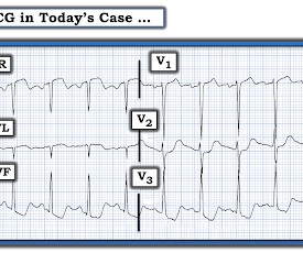
The ECG shows obvious STEMI(+) OMI due to probable proximal LAD occlusion. Epinephrine infusion was begun. The patient in today’s case is a previously healthy 40-something male who contacted EMS due to acute onset crushing chest pain. The below ECG was recorded. Subsequent PCI of the LMCA and LAD was performed.
With ventilations and epinephrine, she regained a pulse. Note that they finally have laid to rest the new or presumably new LBBB as a criteria for STEMI. Note that they finally have laid to rest the new or presumably new LBBB as a criteria for STEMI. A middle-age woman with h/o hypertension was found down by her husband.
There is STE in III and aVF which does not meet STEMI criteria due to insufficient STE in lead aVF. The interventionalists insisted that the ECGs did not meet STEMI criteria and cancelled the activation, stating that they would consider urgent cath after further stabilization. This is an obvious inferoposterior OMI. mm STE with 9.5
Fine ventricular fibrillation She received 2 mg epinephrine, 150 mg amiodarone and underwent chest compressions with the LUCAS device. The last section is a detailed discussion of the research on aVR in both STEMI and NonSTEMI. She arrived in the ED 37 minutes after 911 was called, with continuing CPR. see below).
He underwent CPR, and regained a pulse after epinephrine, with an organized narrow complex rhythm at 140, but still with severe shock. In a series of 18 patients with COVID and ST elevation, 8 were diagnosed with STEMI, 6 of whom had an angiogram and it showed obstructive coronary disease. He was intubated and then went pulseless.
Clinical Course The paramedic activated a “Code STEMI” alert and transported the patient nearly 50 miles to the closest tertiary medical center. 2 The astute paramedic recognized this possibility and announced a CODE STEMI. Look at the aortic outflow tract. What do you see? Answer below in the still shot.
On epinephrine and norepinephrine drips." He had multiple cardiac arrests with ROSC regained each time. Endotracheal tube re-intubation was confirmed multiple times, bilateral breath sounds, yet O2 saturation remained in the 50s and 60s. CT angiogram showed extensive saddle pulmonary embolism.
After epinephrine, atropine, and defibrillation x 2, there was a return of pulses. There is ST depression in II, III, and aVF that is concerning for reciprocal depression from high lateral STEMI in aVL, where there is some ST elevation. She collapsed and 911 was called; she was found pulseless. Exact rhythm during arrest is uncertain.
Resuscitated with chest compressions, epinephrine. A 12-lead was recorded, showing "STEMI," but is unavailable. including epinephrine, and there was ROSC. This is what the providers in the ED understood on patient arrival: Patient called 911 for syncope, then had witnessed PEA arrest after medics arrived. Not a shockable rhythm.
EPINEPHRINE-INUDCED SHOCK: LEFT VENTRICULAR OUTFLOW TRACT OBSTRUCTION ON VASOPRESSORS. m/s)—problematic and elevated > 50 mm Hg (2.5 m/s)—problematic and elevated > 50 mm Hg (2.5 Left ventricular outflow tract gradient variability in hypertrophic cardiomyopathy. Clin Cardiol. 2009;32(7):397-402. doi:10.1002/CLC.20594
In the EMS setting, the most common cardiogenic shock patient is most likely a STEMI. In right-sided shock (inferior STEMIs with +RVI), the LV is underfed due to a poorly functioning RV that can’t pump blood across the pulmonary vasculature. As a result of the increased LVEDP, CPP falls. mcg/kg/min.
We organize all of the trending information in your field so you don't have to. Join 5,000+ users and stay up to date on the latest articles your peers are reading.
You know about us, now we want to get to know you!
Let's personalize your content
Let's get even more personalized
We recognize your account from another site in our network, please click 'Send Email' below to continue with verifying your account and setting a password.


Let's personalize your content