This site uses cookies to improve your experience. To help us insure we adhere to various privacy regulations, please select your country/region of residence. If you do not select a country, we will assume you are from the United States. Select your Cookie Settings or view our Privacy Policy and Terms of Use.
Cookie Settings
Cookies and similar technologies are used on this website for proper function of the website, for tracking performance analytics and for marketing purposes. We and some of our third-party providers may use cookie data for various purposes. Please review the cookie settings below and choose your preference.
Used for the proper function of the website
Used for monitoring website traffic and interactions
Cookie Settings
Cookies and similar technologies are used on this website for proper function of the website, for tracking performance analytics and for marketing purposes. We and some of our third-party providers may use cookie data for various purposes. Please review the cookie settings below and choose your preference.
Strictly Necessary: Used for the proper function of the website
Performance/Analytics: Used for monitoring website traffic and interactions
Traditionally, emergency providers looked for signs of ST-segment elevation myocardial infarction (STEMI) to indicate the need for intervention. Emergency physicians have recognized for some time that there are many occlusions of the coronary arteries that do not present with classic STEMI criteria on the ECG.
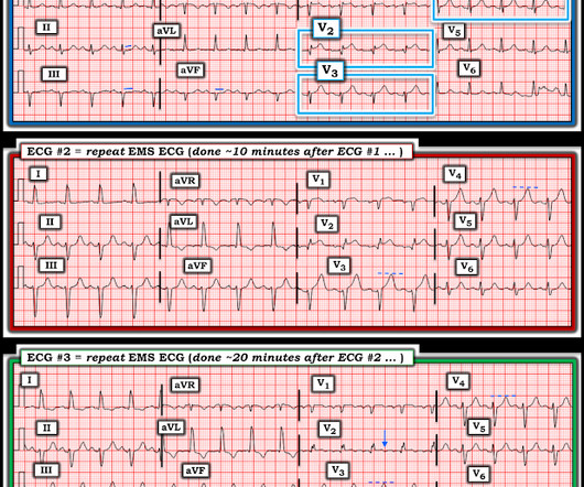
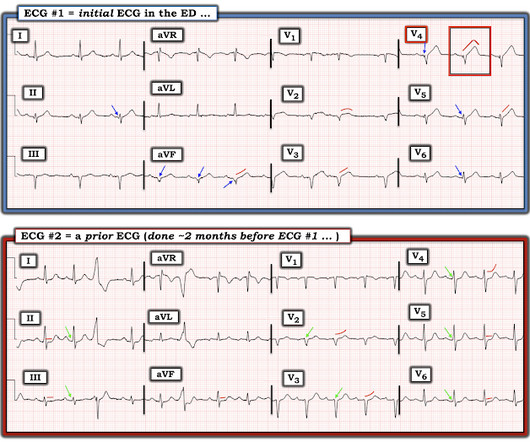
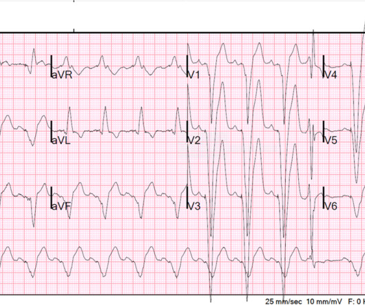
Interpretation : diagnostic of acute anterior OMI with STE less than STEMI criteria in V1-V4, hyperacute T waves in V2-V4, and suspiciously flat isoelectric ST segments in III and aVF suspicious for reciprocal findings. Now it even meets STEMI criteria, and HATWs continue to inflate. So the cath lab was not activated. Ongoing OMI.
The ECG did not meet STEMI criteria, and the final cardiology interpretation was “ST and T wave abnormality, consider anterior ischemia”. There’s only minimal ST elevation in III, which does not meet STEMI criteria of 1mm in two contiguous leads. But STEMI criteria is only 43% sensitive for OMI.[1]
Further information is not available. Despite anticipation by many that the initial post-resuscitation ECG will show an obvious acute infarction — this expected "STEMI picture" is often not seen. Only one hs troponin I was measured on arrival: 323 ng/L Initial echo showed10% EF, diffuse severe hypokinesis.
The biggest problem with STEMI criteria are false negatives – because this costs patient’s myocardium, with greater mortality and morbidity. I sent both ECGs to Dr. Smith, with the only information that these were prior vs new ECG. For this reason, ECGs need first to be interpreted in isolation, and then applied to the patient.
Sent by anonymous, written by Pendell Meyers I received a text with this image and no other information: What do you think? The interventional cardiologist then canceled the activation and returned the patient to the ED without doing an angiogram ("Not a STEMI"). I simply texted back: "Definite posterior OMI."
This ECG was texted to me with the implied question "Is this a STEMI?": I responded that it is unlikely to be a STEMI. Septal STEMI often has ST depression in V5, V6, reciprocal to V1. Then combine with clinical presentation and low pretest probability 2 Saddleback STEMIs A Very Subtle LAD Occlusion.T-wave wave in V1??
Written by Pendell Meyers Both of these cases were sent to me with no information other than adults with acute chest pain. Thus, this does NOT meet STEMI criteria (though, as of 2022, it is a formal "STEMI equivalent", assuming everyone agrees that this is de Winter morphology, for which there is currently no objective definition).
This case was recently posted by Tyron Maartens on Facebook EKG club (he agreed to let me post it here), with the following clinical information: "42 year old male with two weeks of intermittent chest discomfort, awoke 4 hours prior to this ECG with a more severe, heavy chest pain (5/10). Both support acute anterior STEMI. 3.0 = 0.50
So this NSTEMI was likely a STEMI(-)OMI with delayed reperfusion. The patient was admitted as ‘NSTEMI’ which is supposed to represent a non-occlusive MI, but the underlying pathophysiology is analogous to a transient STEMI. See these posts: Chest Pain, ST Elevation, and an Elevated Troponin: Should we Activate the Cath Lab?
He has a history of STEMI and heart failure. I read this blinded, with no clinical information, and read it as inferior OMI. The Queen of Hearts interprets it blinded also (no clinical information and no previous ECGs or serial ECGs). link] Case continued The conventional algorithm diagnosed STEMI and so did the paramedics.
This ECG was texted to me with no clinical information , and I texted back: "RBBB with RVH and inferior-posterior-lateral subacute MI. It is uncommon in the age of reperfusion therapy, as most STEMI get treated reasonably early, before transmural infarct. Most STEMI peak at over 10 ng/mL; most NonSTEMI at less than 10 ng/mL.
This ECG was texted to me with no other information. It does, in fact, the STE meets STEMI criteria since there is 1 mm of in V4 and V5. I assumed the presentation was consistent with acute MI. What did I say? Activate the cath lab." The T-waves in V2-V6 are diagnostic. There is also some non-diagnostic STE in inferior leads.
Jason was very skeptical of STEMI. This also argues against STEMI. But knowing this information ( as described above ) — allows us to rapidly identify the ECG in Figure-1 as one more manifestation of this patient's baseline ECG. He complained of 3 days of diarrhea and abdominal pain. What do you think? Jason, I agree.
There’s inferior ST depression which is reciprocal to subtle lateral convex ST elevation, and the precordial T waves are subtly hyperacute – all concerning for STEMI(-)OMI of proximal LAD. There’s ST elevation I/aVL/V2 that meet STEMI criteria. This is obvious STEMI(+)OMI of proximal LAD. Non-STEMI or STEMI(-)OMI?
Recall from this post referencing this study that "reciprocal STD in aVL is highly sensitive for inferior OMI (far better than STEMI criteria) and excludes pericarditis, but is not specific for OMI." What would you do at this time with this information? But pain is an important signal in MI and informs the clinician of the urgency.
Pulsara , a communication app that’s available on iOS, Android and web browsers, allows members of the department to share patient information with every member of the care team through an intuitive platform. Using Pulsara daily has allowed those at CSFD to build muscle memory, making emergency management that much easier.
This patient could have very easily been overlooked, both because the ECG was STEMI negative and because the Q waves were attributed to an “old infarct”. Fortunately, Dr. Cho was not looking for STEMI ECG criteria but for an acute coronary occlusion. OMI or STEMI? As cardiology documented, “possible STEMI.
Are Some Cardiologists Really Limited by Strict Adherence to STEMI millimeter criteria? I was texted these ECGs by a recent residency graduate after they had all been recorded, along with the following clinical information: A 50-something with no cardiac history, but with h/o Diabetes, was doing physical work when he collapsed.
He reports that this chest pain feels different than prior chest pain when he had his STEMI/OMI, but is unable to further describe chest pain. I sent it to 5 of my OMI friends without any clinical information or outcome and all 5 independently responded with exactly the same diagnosis: "reperfused inferior OMI".
The ECG shows an inferior ST-Elevated Myocardial Infarction (STEMI). They felt this would help inform guideline writers on making recommendations in this area. His vitals are: heart rate of 72 beats per minute, blood pressure of 150/90, respiratory rate of 14 breaths per minute and oxygen saturation of 93%.
The chest pain started about 24 hours ago, but there was no detailed information available about whether his pain had come and gone, or what prompted him to be evaluated 24 hours after onset. As most would agree, this ECG shows highly specific findings of anterolateral OMI, even with STEMI criteria in this case. Learning Points: 1.
A prehospital “STEMI” activation was called on a 75 year old male ( Patient 1 ) with a history of hyperlipidemia and LAD and Cx OMI with stent placement. The two cases were considered: Patient 1 was recognized by the ED provider and the cardiologist as having resolved “STEMI”. He wrote most of it and I (Smith) edited.
Any ST Depression Maximal in V1-V4 is OMI until proven otherwise I sent this ECG with no information to Pendell. Here it is: Obvious Inferior Posterior STEMI (+) OMI. Initial troponin was: 3 ng/L We showed that the first troponin in acute STEMI is often negative in at least 27%. We send each other EKG by the dozens every day.
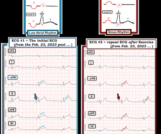
In the available view of the sinus rhythm, we see normal variant STE which probably meets STEMI criteria in V4 and V5. In other words, the inferior "ST elevation" is due to the abnormal rhythm, and does not signify OMI or STEMI in any way. Admittedly — we often are not privileged to see this information.
A post-arrest ECG doesn’t show any signs of STEMI. For more information on the fragility index (FI) click on this LINK. The patient is in ventricular fibrillation, and you achieve return of spontaneous circulation (ROSC) on the second shock. The patient is still unconscious. Are we supposed to be starting hypothermia?”
The ANSWER: At this point in the case — I was provided with 2 additional pieces of information: #1 Informational: It turns out that the patient in today's case was critically ill with multisystem problems. Synchronized cardioversion @200J was attempted twice on the rhythm in ECG #1 — but this had no effect on the rhythm. #2
Optical coherence tomography, due to its high resolution, may provide additional information [ 10,13 ]. STEMI MINOCA versus NSTEMI MINOCA STEMI occurs in the presence of transmural ischaemia due to transient or persistent complete occlusion of the infarct-related coronary artery. From Gue at al. Circulation. 2017;135(16):1490–3.
He sent it to me with no other information and I wrote back "100% diagnostic of LBBB with inferior-posterior-lateral OMI" There is atrial paced rhythm with Left Bundle Branch Block (LBBB). Most large STEMI have peak troponin I in the 20.0 This ECG was recorded and was reviewed remotely by a cardiologist: What do you think?
The receiving emergency physician consulted with interventional cardiology who stated there was no STEMI. Learning points: Both patients and other medical providers can report confusing and often contradictory information that obfuscates the diagnosis (in this case, WPW). Is there STEMI? The patient continued having chest pain.
Only very slight STE which does not meet STEMI criteria at this time. I am immediately worried that this OMI will not be understood, for many reasons including lack of sufficient STE for STEMI criteria, as well as the common misunderstanding of "no reciprocal findings" which is very common with this particular pattern.
Inspecting the morphology can give us more information about the source of the escape rhythm. This is documented as a STEMI in the clinical notes and in the cath report, but certainly does not meet STEMI criteria and is therefore an NSTEMI by definition. Most STEMI have peak cTnI greater than 10.0. ng/mL (ref. <
The prehospital and ED computer interpretation was inferior STEMI: There’s normal sinus rhythm, first degree AV block and RBBB, normal axis and normal voltages. The paramedic notes called STEMI into question: “EMS disagree with monitor for STEMI callout. Vitals were normal except for oxygen saturation of 94%. Vitals were normal.
Lets look at a few and make an informed decision. His initial EKG is the following: What do you think? Are there any meds you can give? Should you? Discussion To be fair, that question has been the primary focus of multiple review studies and trials over the years.
I texted this ECG with no information to Dr. Smith, who immediately said: "If CP, then anterior OMI until proven otherwise." Post Cath ECG: Obviously completing MI with LVA morphology, and STE that meets STEMI criteria (but pt is still diagnosed as "NSTEMI"). No TIMI flow was listed in the report.
EMS recorded this ECG during active symptoms and transmitted it to the ED: I had no information when I was shown the ECG. I believe there is not quite enough STE for formal STEMI criteria, but some might measure 1.0 I believe there is not quite enough STE for formal STEMI criteria, but some might measure 1.0 I said "Not OMI.
As for precordial leads: this is from my files and I don't have all the information, but it is clearly a right sided ECG (QRS in V5, V6 is inverted from QRS in I, aVL) What else do you see? The cath lab was activated for STEMI. So, in retrospect, the first patient probably did not have STEMI at all. There was no MI. Lessons : 1.
But because there was no new ST elevation, the ECG was signed off as “STEMI negative” and the patient waited to be seen. But the ECG still doesn’t meet STEMI criteria. It was therefore interpreted as “no STEMI” and the patient was treated with dual anti-platelets and referred to cardiology as “NSTEMI.” the cardiologist 5.
This is a particularly informative link: 2 Examples of Posterior Reperfusion T-waves So one might think that, with active pain, there is anterior OMI. Is she seeing anterior hyperacute T-waves, or does she see Posterior Reperfusion T-waves? I think the ECG is normal. Therefore, we activate the Cath Lab.
We don’t know any of this information unfortunately and all are key in patient selection The median lactate level before revascularization was 6.9mmol/L (Range 4.6 Control: 53.4% D ECLS: 18.2% Control 8.7% Control 38.0% Control: 49.0% RR 0.98; 95% CI 0.80 to 1.19; p = 0.81 vs 13.9% (RR 0.58; 95% CI 0.33 vs 22.6% (RR 1.03; 95% CI 0.88
You can read this post here, or watch a video presentation of it: [link] I was handed this ECG, without any clinical information, while on my way to see another patient: There is sinus rhythm. If there are no changes in aVL, it is highly unlikely to be inferior STEMI. Would you be certain that it is not STEMI? mm in V3, 2.5
I sent the ECG to Dr. Meyers without any other information. Now let’s compare this with the existing paradigm to identify multiple preventable delays to reperfusion, which can be improved through the paradigm shift from STEMI to OMI. But STEMI criteria has poor sensitivity for acute coronary occlusion.
is very specific for STEMI , and there is some evidence, as well as rationale, that a paced rhythm behaves similarly. Here is one case of anterior STEMI in a paced rhythm. Here is a case of lateral STEMI in a paced rhythm. Use all the information at your disposal to assess the situation. Lesson : 1.
I want all to know that, with the right mind preparation, and the use of the early repol/LAD occlusion formula, extremely subtle coronary occlusion can be detected prospectively, with no other information than the ECG. His ECG was repeated at this point: This shows a well developed anterior STEMI. The peak troponin I was over 100.
We organize all of the trending information in your field so you don't have to. Join 5,000+ users and stay up to date on the latest articles your peers are reading.
You know about us, now we want to get to know you!
Let's personalize your content
Let's get even more personalized
We recognize your account from another site in our network, please click 'Send Email' below to continue with verifying your account and setting a password.


Let's personalize your content