This site uses cookies to improve your experience. To help us insure we adhere to various privacy regulations, please select your country/region of residence. If you do not select a country, we will assume you are from the United States. Select your Cookie Settings or view our Privacy Policy and Terms of Use.
Cookie Settings
Cookies and similar technologies are used on this website for proper function of the website, for tracking performance analytics and for marketing purposes. We and some of our third-party providers may use cookie data for various purposes. Please review the cookie settings below and choose your preference.
Used for the proper function of the website
Used for monitoring website traffic and interactions
Cookie Settings
Cookies and similar technologies are used on this website for proper function of the website, for tracking performance analytics and for marketing purposes. We and some of our third-party providers may use cookie data for various purposes. Please review the cookie settings below and choose your preference.
Strictly Necessary: Used for the proper function of the website
Performance/Analytics: Used for monitoring website traffic and interactions
When I started as a paramedic in Hartford in January of 1995, I was given a 100-page protocol book to memorize. For instance, there was no protocol for stroke or STEMI. For instance, there was no protocol for stroke or STEMI. Paramedics didnt do 12-lead ECGs then. Looking through the book today, it is truly an antiquated.
The paramedic called the EM physician ahead of arrival and discussed the case and ECGs, and both agreed upon activating "Code STEMI" (even though of course it is not STEMI by definition), so that the acute LAD occlusion could be treated as fast as possible. So the cath lab was activated. Long term outcome is unavailable.
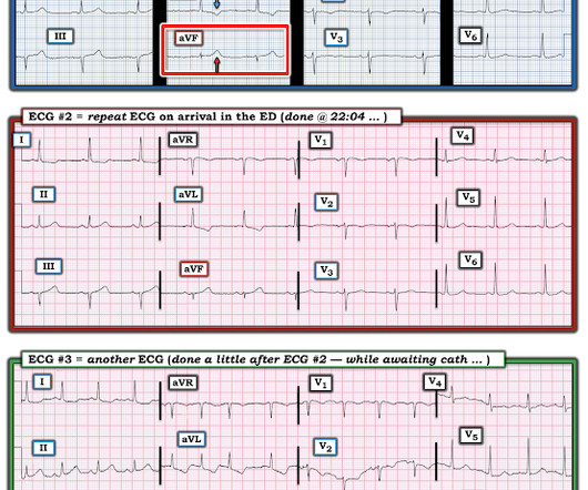
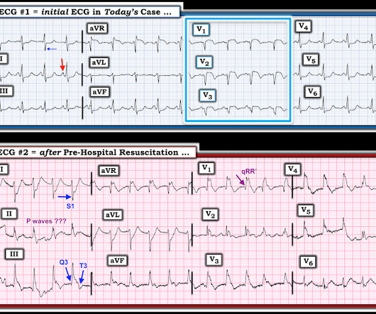
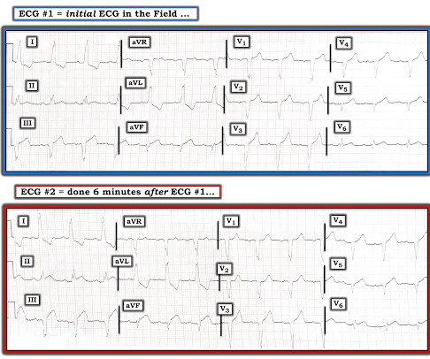
Another ECG was recorded while awaiting the cath team: Now there is STEMI Let's look at that first (prehospital ECG) again: Very subtle! A prehospital activation would have save a lot of time and would have been possible if the paramedics were using the Queen of Hearts PMCardio AI app. We activated the cath lab.
Serial ECGs demonstrated dynamic changes diagnostic of ACS (transient STEMI) 4. Finally, Transient STEMI should be taken emergently to the cath lab. Normalization of Diagnostic For STEMI Prehospital ECG with Nitroglycerin Therapy. If the initial ECG was diagnostic for STEMI the paramedic called to mobilize the reperfusion team.
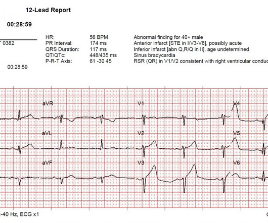
Below is the first ECG recorded by paramedics after 2 hours of chest pain, interpreted by the machine as “possible inferior ischemia”. While STEMI negative, the ECG is diagnostic of proximal LAD occlusion. In isolation this ECG does not show OMI, but following the paramedic ECGs this indicates spontaneous LAD reperfusion.
In this ECG Cases blog we review 8 cases of patients with prehospital ECGs and explore prehospital ECGs for diagnosing STEMI, Occlusion MI, false STEMI, code STEMI, dynamic ischemic changes, truncated voltages. Can you avoid the pitfalls and spot the pearls that help to make the diagnosis?
So while there’s no diagnostic STEMI criteria, there are multiple ischemic abnormalities in 11/12 leads involving QRS, ST and T waves, which are diagnostic of a proximal LAD occlusion. First trop was 7,000ng/L (normal 25% of ‘Non-STEMI’ patients with delayed angiography have the exact same pathology of acute coronary occlusion.
David Didlake Acute Care Nurse Practitioner Firefighter / Paramedic (Ret) @DidlakeDW Expert contribution by Dr Robert Herman @RobertHermanMD @PowerfulMedical (Chief Medical Officer) An adult male called 911 for new-onset epigastric burning. To which the lead paramedic replied, “Not cardiac; his symptoms are atypical. Is this OMI?
David Didlake Acute Care Nurse Practitioner Firefighter / Paramedic @DidlakeDW A 50 y/o Male was taking his dog for a leisurely stroll through the park when he suddenly experienced new onset chest discomfort. it has been subsequently deemed a STEMI-equivalent.
The ECG is determined to be non-diagnostic by the treating paramedic. The treating paramedic withholds aspirin. At the hospital a 12-lead ECG is recorded within 10 minutes and read by the attending physician, who activates the “Code STEMI” protocol. The paramedic, feeling a bit sheepish, asked me to review the case.
Paramedics provided another 3 sprays of nitro, and 6mg of morphine, which reduced but did not resolve the pain. I sent this to the Queen of Hearts So the ECG is both STEMI negative and has no subtle diagnostic signs of occlusion. of such ‘high risk Non-STEMI’ patients get angiography within 2 hours.[2] But only 6.4%
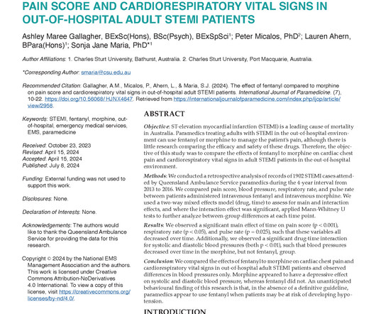
Objective: ST-elevation myocardial infarction (STEMI) is a leading cause of mortality in Australia. Paramedics treating adults with STEMI in the out-of-hospital environment can use fentanyl or morphine to manage the patient’s pain, although there is little research comparing the efficacy and safety of these drugs.
Written by Jesse McLaren Two 70 year olds had acute chest pain with nausea and shortness of breath, and called paramedics. There’s inferior ST depression which is reciprocal to subtle lateral convex ST elevation, and the precordial T waves are subtly hyperacute – all concerning for STEMI(-)OMI of proximal LAD. Who needs the cath lab?
He was a paramedic at the time. Such proficient interpreters include health care assistants and EKG technicians. Pendell Meyers had not started medical school by summer of 2012, but he had read every one of my blog posts over the preceding 4 years. By the summer of 2012, he could read an ECG for OMI better than any doctor I knew.
Jason was very skeptical of STEMI. This also argues against STEMI. Outcome "I later found out that this is a patient who regularly calls paramedics to c/o chest pains and he had fooled many of them. He complained of 3 days of diarrhea and abdominal pain. What do you think? Jason, I agree. There is high R-wave voltage.
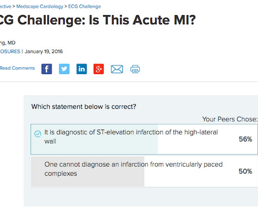
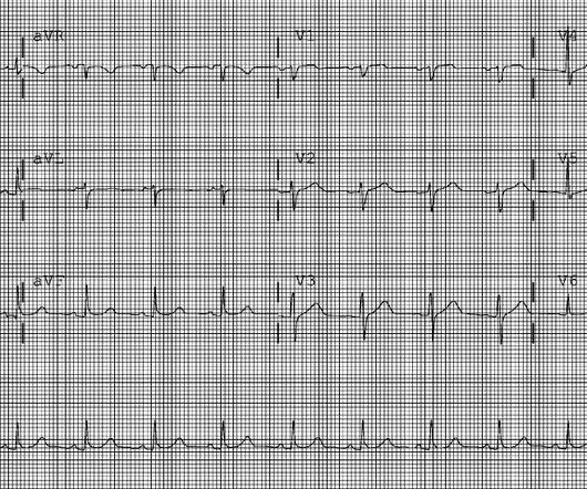
Notice on the right side of the image how the algorithm correctly measures STE sufficient in V1 and V2 to meet STEMI criteria in a man older than age 40. As most would agree, this ECG shows highly specific findings of anterolateral OMI, even with STEMI criteria in this case. Thus, this is obvious STEMI(+) OMI until proven otherwise.
Patient 1 : a 75 year old called paramedics with one day of left shoulder pain which migrated to the central chest, which was worse with deep breaths. The prehospital and ED computer interpretation was inferior STEMI: There’s normal sinus rhythm, first degree AV block and RBBB, normal axis and normal voltages. Vitals were normal.
The paramedics achieve return of spontaneous circulation (ROSC) after CPR, advanced cardiac life support (ALCS), and Intubation. There is evidence that taking those patients with ROSC and EKG showing STEMI directly for angiography +/- angioplasty is associated with positive patient-oriented outcomes.
Madden, Paramedic. There is mixed overlap of ST-segment elevation (STE), ST-segment depression (STD), Hyperacute T waves (HATW), and deWinter pattern (which the ACC regards as a STEMI-equivalent but is better suited under the blanket of OMI). Let's revisit the deWinter occlusion provided by Paramedic Madden.
David Didlake Firefighter / Paramedic Acute Care Nurse Practitioner @DidlakeDW Peer review by Dr. Stephen Smith @smithECGblog I was reviewing ECG’s in our LifeNet database and happened upon this one without any knowledge of clinical circumstances. 1] Here is the admitting ED ECG after cancellation of Code STEMI. 1] Driver, B.
David Didlake Firefighter / Paramedic Acute Care Nurse Practitioner @DidlakeDW Peer review and commentary by Dr. Steve Smith [link] @SmithECGblog It is early-summer, approximately 1330 hours, no cloud cover overhead, and 86 degrees with high humidity. As it currently stands, an ST/S ratio >15% should raise awareness for new anterior STEMI.
Prehospital – hospital interoperability As previously mentioned, the EMS industry is evolving and with it, so is the job of the paramedic. For Eagle County Paramedic Services, turning to MIH was integral to helping them provide services for their underserved population while also saving millions in costs.
He has a history of STEMI and heart failure. link] Case continued The conventional algorithm diagnosed STEMI and so did the paramedics. A Coronary angiogram from 8 years prior revealed that he had had an inferior posterior STEMI at the time due to 100% occlusion of the proximal RCA. He was belted and it was low speed.
David Didlake Firefighter / Paramedic Acute Care Nurse Practitioner @DidlakeDW Peer review provided by Dr. Steve Smith [link] @SmithECGBlog An adult female called 911 for chest discomfort and difficulty breathing. Then, three minutes later… Crews activated STEMI as she deteriorated into PEA arrest.
David Didlake Firefighter / Paramedic Acute Care Nurse Practitioner @DidlakeDW Peer review provided by Dr. Steve Smith @SmithECGblog I was conducting QA/QI on two very recent cases and was struck by the uniqueness of both. A prehospital STEMI activation was transmitted to the closest PCI center, and 324mg ASA was administered.
He called 911 and paramedics recorded a prehospital 12 lead ECG which showed a clear inferior STEMI (not shown, tracing could not be found). The cath lab was activated by the paramedics. Objectives : To find the incidence of any rSTD or T-wave inversion (TWI) in angiographically proven inferior STEMI. There is 0.5
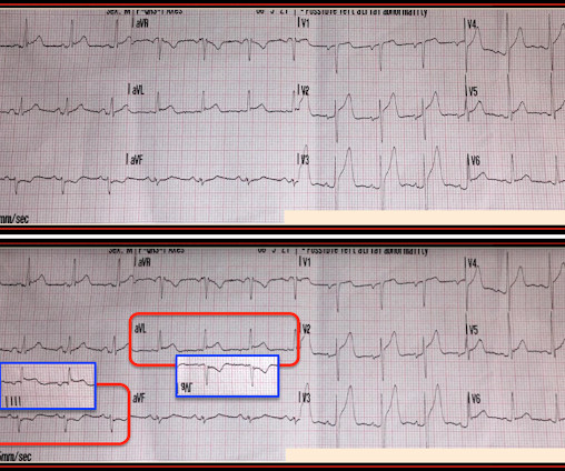
Acute anterior STEMI tends to be a more difficult ECG diagnosis than acute inferior STEMI. That’s because with acute inferior STEMI there’s almost always a downsloping ST-segment in lead aVL to help shore up the diagnosis. Once again, this acute anterior STEMI “crosses over” to the high lateral leads.
David Didlake Firefighter / Paramedic Acute Care Nurse Practitioner @DidlakeDW Expert commentary provided by Dr. Ken Grauer CASE 1 An 82 y/o Male called 911 for sudden onset dizziness while at rest. ASA 324mg was administered while a STEMI activation was simultaneously transmitted to the nearest PCI center. Attached is the first ECG.
RBBB in acute STEMI has a very high mortality. The paramedics activated the cath lab from the field. Thus, there is right bundle branch block, which should never (unlike Left BBB) have any ST elevation. But here there is a large degree of ST elevation in V2-V6, I, and aVL.
She was diagnosed with a Non-STEMI and kept overnight for a next day angiogram. Paramedics found her semi-conscious, pale, cool, diaphoretic, tachypneic, very hypotensive. Medics recorded the above ECG and called a STEMI alert. Her troponin I returned at 900 ng/L. Patient was given aspirin, sublingual nitro as well as heparin.
This case was sent by Lou B, a paramedic and RN. Here it is: The computer reads STEMI What do you think? More from the medic: "LifePak 15 interpretation was STEMI. My response: "I think it is very worrisome for STEMI." It meets STEMI criteria even for a male under age 40, with STE 2.84 Pattern looked to be BER.
The paramedic recorded a series of ECGs; the initial ECG is representative here: Computer read: “ Normal ECG ” What do you think? The paramedic interpreted this as a STEMI. Can you employ the Subtle Anterior STEMI calculator ? It is not yet available, but this is your way to get on the list. or LAD occlusion?
Her initial 12-lead ECG that was obtained by paramedics in the field is shown in Figure-1. Figure-1: The initial ECG in todays case, obtained by paramedics in the field. ( On seeing both of the tracings in Figure-2 the paramedic team and the ED physician activated the after-hours cath lab. Should you activate the cath lab?
Jason was very skeptical of STEMI. This also argues against STEMI. Outcome "I later found out that this is a patient who regularly calls paramedics to c/o chest pains and he had fooled many of them. Look for old ECGs Do serial ECGs Do echocardiography June 17, 2016 Anterior STEMI? What do you think? Jason, I agree.
This was shown to me by a very astute Hennepin paramedic. Although this comes from a Hennepin paramedic, the patient was not brought to Hennepin County Medical Center. It is important for cardiologists to realize that a paramedic may see something they do not. of this post. This is my reponse. This is not tribalism.
My most talented blog readers are paramedics because they have to put themselves on the line every time they activate the cath lab. Trop T now very high, well into the range one sees with a STEMI; very unusual in type II MI. The minute this medical student saw the first ECG, he knew the diagnosis without any further information.
This case was provided by Spencer Schwartz, an outstanding paramedic at Hennepin EMS who is on Hennepin EMS's specialized "P3" team, a team that receives extra training in advanced procedures such as RSI, thoracostomy, vasopressors, and prehospital ultrasound. Learning Points: 1. Learn to Recognize Hyperacute T-waves 2. From Gue at al.
The paramedics found the patient with ROSC and a GCS 7, and an ECG showing LBBB with possible lateral ST elevation. The patient was brought to the ED as a possible Code STEMI and was seen directly by cardiology. On arrival, GCS was 13 and the patient complained of ongoing chest pain.
When the paramedics arrived, they obtained a 12 lead ECG and confirmed the unstable vital signs. There is an obvious inferior STEMI, but what else? Besides the obvious inferior STEMI, there is across the precordial leads also, especially in V1. This STE is diagnostic of Right Ventricular STEMI (RV MI).
Clinical Course The paramedic activated a “Code STEMI” alert and transported the patient nearly 50 miles to the closest tertiary medical center. 2 The astute paramedic recognized this possibility and announced a CODE STEMI. Look at the aortic outflow tract. What do you see? Answer below in the still shot.
We have a large number of graduate paramedics starting with Ambulance Victoria this year, so it’s probably a good time to revisit a topic that seems to receive surprisingly little attention in an industry that relies so heavily upon it. Sometimes I just have to scratch the itch and have a rant. Learn it, use it. Listen more.
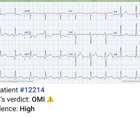
Here’s the paramedic ECG (digitized by PMcardio). STEMI negative : the EMS automated interpretation read, “STEMI negative. But the latest ACC consensus on the evaluation of chest pain in the ED warns that “STEMI criteria will miss a significant minority of patients with acute coronary occlusion.”[1] What do you think?
For Eagle County Paramedic Services, turning to mobile integrated healthcare was integral to helping them provide services for their underserved while also saving millions in costs. Supporting and advocating for mental health Mental health calls are increasing.
Here is the written paramedic report available after all the events were over: Patient was seen by witnesses to become unresponsive. A 12-lead was recorded, showing "STEMI," but is unavailable. Moreover, when someone has immediate resuscitation of an arrest witnessed by paramedics, they rarely have a GCS of 3 (deep coma).
We organize all of the trending information in your field so you don't have to. Join 5,000+ users and stay up to date on the latest articles your peers are reading.
You know about us, now we want to get to know you!
Let's personalize your content
Let's get even more personalized
We recognize your account from another site in our network, please click 'Send Email' below to continue with verifying your account and setting a password.


Let's personalize your content